MEDRADSC 2L03 Lecture Notes - Lecture 1: Abdominal Aorta, Adventitia, Aorta

24 Sep 2018
School
Department
Course
Professor
Document Summary
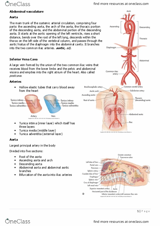
They have 3 coats, or tunics: adventitia, fibrous, outermost, media, muscle and elastic, middle layer. The abdominal aorta is the blood supply to the abdominopelvic viscera, walls, and lower extremities. Divided into segments: ascending, arch, descending, thoracic, abdominal. Becomes the abdominal after it pierces the diaphragm. Branches: celiac trunk, left gastric, splenic, common hepatic, gastroduodenal, right gastric, cystic, sma, renals, left and right. Top left: the ct is coming out of the aorta. Top right: sma coming out just after the ct (a lot more slanted and longer) Bottom left: left and right common iliac artery. This is the aorta at the right crus. This is the required image for test. You can see the ct coming off the top of the aorta, which then splits into the sphlenic artery an \d the common hepatic artery. Celiac trunk can be seen a bit better, seagles wings. To the right is the splenic artery, and to the left is the cha.