MEDRADSC 2I03 Lecture Notes - Lecture 12: Costodiaphragmatic Recess, Airway Obstruction, Chest Tube

28 Jul 2019
School
Department
Course
Professor
Document Summary
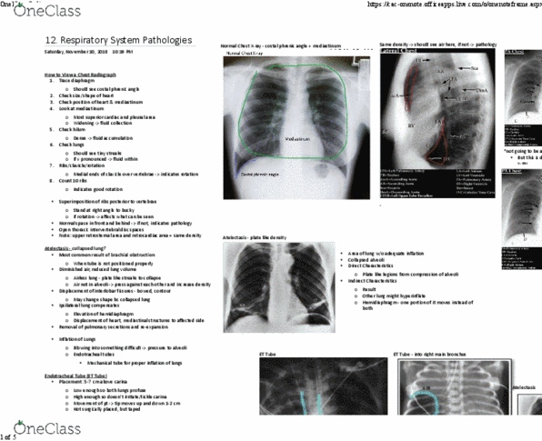
How to view a chest radiograph: pathology of the respiratory system. Haven"t clipped anything (apices to the base of the lungs) Trace diaphragm: always see the costophrenic angle (dip on lateral edges of the diaphragm; want to see it) Check position of heart & mediastinum: mediastinal shift = huge indication that something is wrong. Look at mediastinum: most superior aspect of the cardiac & pleural area; on top of the heart; indicative of fluid collection. Check hilum: see main vessels; if it looks dense = fluid accumulation in vessels. Check lungs: if streaks are very pronounced = lots of fluid; want to see small streaks. Ribs/clavicle/rotation: if the clavicle is over top of the spine, you know patient is rotated. If the patient doesn"t take a good enough breathe in, pathologies can be missed. Normal space b/n heart & vertebrae and normal space b/n heart & sternum.