BIOLOGY 2B03 Lecture Notes - Lecture 7: Osmosis, Lipid Raft, Acylation

6 Jun 2018
School
Department
Course
Professor

Biomembranes and Cell Architecture: Biomembranes
Biomembranes
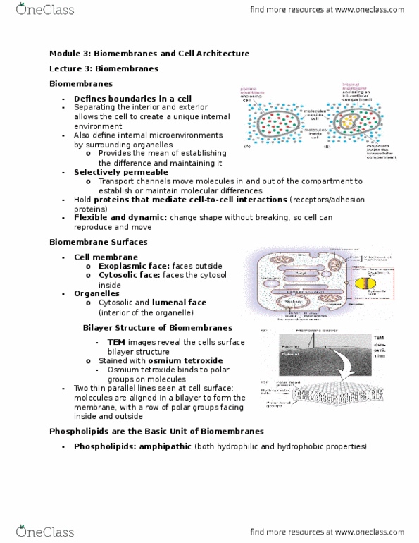
define compartment boundaries •
are selectively permeable •
transport channels move molecules in and out of compartment to maintain molecular differences ◦
very few can move freely across membranes ◦
flexibility/dynamics allow cell to bend/change shape without breaking •
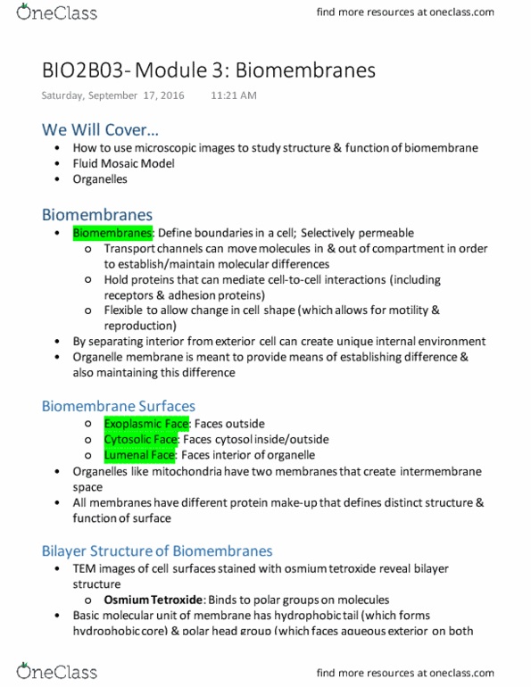
bio membrane surfaces: •
cytosolic ◦
extoplasmic ◦
lumenal ◦
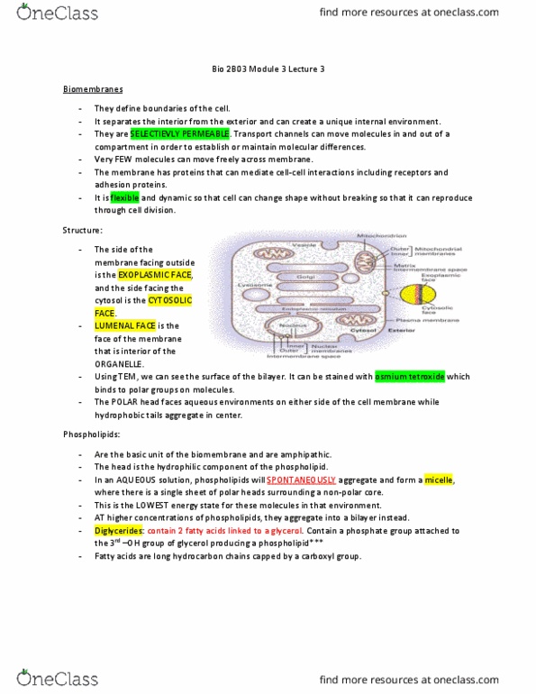
bilayer structure of biomembranes: •
TEM images can reveal its bilayer structure ◦
Phospholipids
basic unit of biomembranes •
amphipathic - hydrophilic and hydrophobic •
phospholipids arrange themselves in aqueous solution to form bubble-like structure called a micelle •
lowest energy state in this env't ◦
at higher concentration of phospholipids, can spontaneously assemble a bilayer •
diglycerides contain 2 fatty acids linked to glycerol •
contain phosphate group attached to third -OH group of glycerol which produces a phospholipid ◦
phosphate group is hydrophilic - fatty acid is hydrophobic ◦
all biomembranes have same basic structure, but each has distinctive activities due to its complement of proteins •
Integral membrane proteins: embedded in hydrophobic core of bilayer ◦
Peripheral membrane proteins: associated with one surface ◦
Fluidity
fluidity allows membranes to fuse with each other •
can deform without tearing and change shape •
allows lateral movement within bio membrane •
each layer of phospholipid membrane called a leaflet •
has different properties based on the embedded proteins ◦
Fluid Mosaic Model
proposed by S.J Singer and G Nicolson in 1972 •
describes structural features of biological membranes •
plasma membrane described as fluid because components + phospholipids + membrane proteins move laterally •
through membrane meaning it is not solid, but more 'fluid'
described as 'mosaic' because made of many different ◦
macromolecules
FRAP: Measuring Fluidity
microscopic labelling technique - to track and measure fluidity of •
proteins in membrane
stands for Fluorescence Recovery After Photobleaching •
diagram shows experiment where single kind of membrane •
protein (orange) is distributed in cell membrane
fluorescent tags (antibody) added to tagged via GFP ◦
fluorescent molecule excited by UV light (not too much ◦
otherwise get bleached/saturated and not fluoresce)
Document Summary
Integral membrane proteins: embedded in hydrophobic core of bilayer. Fluidity fluidity allows membranes to fuse with each other can deform without tearing and change shape allows lateral movement within bio membrane each layer of phospholipid membrane called a lea et has different properties based on the embedded proteins. Single-pass membrane proteins protein with single hydrophobic alpha helix spans phospholid bilayers leaving domains on exterior and interior surfaces of membrane. Is a homodimer and is a single pass transmembrane protein. Multi-pass transmembrane proteins: pass through membranes many times uses 7 membrane spanning domain with 7 alpha helices that interact with each other to form transmembrane domain. Bacteriorhodopsin - includes singling receptors such as g-protein coupled receptors. Beta-barrel: beta barrel formed from 16 beta strands exterior of barrel is hydrophobic so can interact with membrane and interior is hydrophilic forms hydrophilic pore through hydrophobic membrane. Channels collection of alpha helices exterior is hydrophobic and interior is hydrophilic.