PSYC 444 Lecture Notes - Lecture 3: Slow-Wave Sleep, Rapid Eye Movement Sleep, Ultradian Rhythm

14 May 2018
School
Department
Course
Professor

PSYC 444 – LECTURE 3
SLEEP ARCHITECTURE: HYPNOGRAM
Progression of sleep stages throughout the night can be seen on the hypnogram
By looking at the waveform, able to determine which stage of sleep the person is in
WAKING EEG
Fast, desynchronized activity
• EOG: eye movements
• EMG: muscle activity of the chin
• EEG: brain activity
It is difficult to measure activity when awake due to many artifacts, such as minor movements like swallowing
WAKING EEG: EYES CLOSED
Smaller waves do not necessarily mean less activity but could be an indication of desynchronized activity.
NREM 2
Desynchronized, mainly theta activity characterized by k-complex and sleep spindles
SLOW WAVE SLEEP (SWS)
Really slow delta activity: low amplitude and low frequency; about 1Hz
Seen more often in younger individuals than old due to more consolidated sleep
REM
It is difficult to distinguish REM from awake state without using an EEG
• EMG is placed on chin to measure muscle atonia, one indicator of REM sleep
• Muscle atonia is paralysis of the body to prevent acting out of dreams
• Loss of muscle atonia during sleep is a characteristic of REM sleep disorder
3 requirements of REM sleep: rapid eye movement, muscle atonia, and mixed wave characteristics
2 parts of REM sleep:
1. Phasic: REM
2. Tonia: anything in between
If you wake someone from REM sleep, there is a high chance that they were immersed in an engaging and realistic dream
• There is a lot of mental activity during REM sleep
EEG of REM sleep is desynchronized with low amplitude
• Rapid eye movements are sharp and perpendicular (very different from slow rolling eye movement)
SLEEP MICROARCHITECTURE
In addition to sleep stages and EEG frequencies, researchers pay special attention to phasic events during sleep. Some of these are:
• Sleep spindles and K-complexes
• Delta waves
• Rapid eye movements
In addition to core measurements, micro-events are looked at to determine the relationship with phenomena such as nightmares, memory, cognitive decline, etc.
REM and nREM cycles are interspersed with awakening and fluctuations from light to deep sleep
• Sleep is more fragmented in the elderly
• The key is proportion of deep and light sleep
(left) Sleep cycles are predictable but they are not equally distributed throughout the night
(right) Sleep deprived participants in the lab (grey: nREM; black: REM)
• Circadian rhythm keeps alternating but sleep debt is continuously accumulated
• Slow wave sleep takes precedence: recover nREM sleep first, then returns to REM-nREM pattern
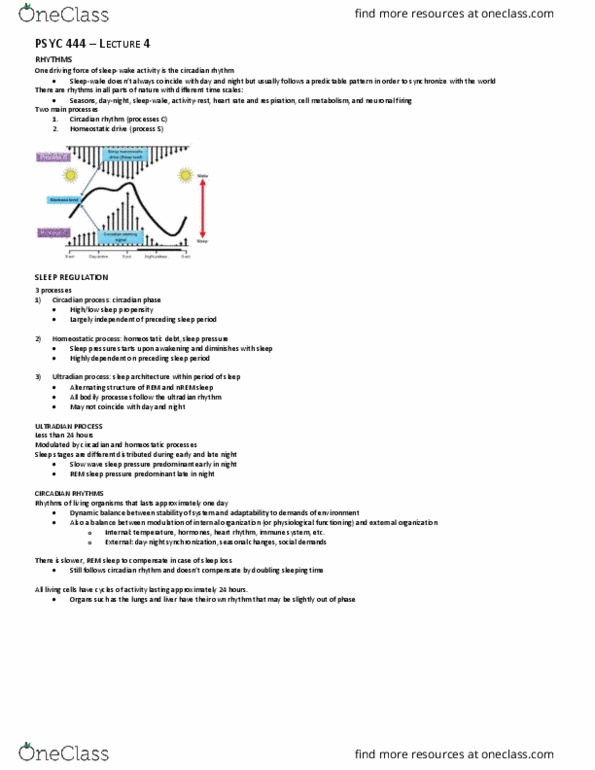
One motor of sleep regulation is the ultradian rhythm (less than 24 hours)
• The ultradian rhythm differs depending on the organ
find more resources at oneclass.com
find more resources at oneclass.com

NEURAL CONTROL OF SLEEP
Wakefulness and sleep are often contrasted as two opposite states of consciousness
• Sleep: interaction between circadian and homeostatic processes
• In reality, sleep is not a state but a process, alternating between nREM-REM cycles
• Sleep and wake are determined by a coordination of two opposing (neurobiological) systems
o Wake-promoting, arousal system
o Sleep-promoting system
Most sleep research involves animal models; little is known about sleep in humans
TRANSECTION STUDIES
Lesions were made in cat brains to determine structures involved in sleep regulation
Cerveau isolè: isolated forebrain (neocortex)
Immediately after midbrain transection, forebrain shows continuous slow waves on EEG
• Structures below the forebrain are likely to be involved in generating sleep signals
Mid-pontine lesions: predominance of arousal/wake
• Neuronal groups localized between the mid-pons and upper midbrain are important for generating a waking-like state
After about a week, the whole brain starts to show circadian rhythms again
• The animal recalibrates, despite lesions to areas important for transmission of sleep signals
• There is no single system that tells the brain to sleep or wake up, but a distributed network that works in concert
(above) creation of artificial lesions
1. Brain is cut from the spinal cord
• Hard to study anything because the animal becomes very unwell
2. Reticular formation plays an important role in general control of a vigilant state
3. Behind pons, the isolated forebrain has effects on all aspects of sleep. It is the center of sleep pattern alternations
OTHER BRAIN AREAS INVOLVED IN SLEEP GENERATION
Thalamus: critical role in regulating EEG patterns
• Wake and sleep promoting areas
• Involved in almost all biological functions
• Determines stage of sleep
Hypothalamus: sleep/wake switch-like mechanism
Lower brainstem: sleep active neurons in the medulla
Many structures throughout the brain are involved in sleep-wake regulation
• Global coordinated process
AROUSAL SYSTEMS OF THE BRAIN
Not a brain area but networks involving many brain areas and neurotransmitters
• Arousal is a global process, involving neurons with long axons with ascending and descending projections
• Damage to one system will result in another pathway compensating for it
o Brain plasticity
find more resources at oneclass.com
find more resources at oneclass.com
Document Summary
Progression of sleep stages throughout the night can be seen on the hypnogram. By looking at the waveform, able to determine which stage of sleep the person is in. It is difficult to measure activity when awake due to many artifacts, such as minor movements like swallowing. Smaller waves do not necessarily mean less activity but could be an indication of desynchronized activity. Desynchronized, mainly theta activity characterized by k-complex and sleep spindles. Really slow delta activity: low amplitude and low frequency; about 1hz. Seen more often in younger individuals than old due to more consolidated sleep. It is difficult to distinguish rem from awake state without using an eeg. Emg is placed on chin to measure muscle atonia, one indicator of rem sleep: muscle atonia is paralysis of the body to prevent acting out of dreams. Loss of muscle atonia during sleep is a characteristic of rem sleep disorder.