BIOL 200 Lecture Notes - Lecture 19: Polytene Chromosome, Karyotype, Fluorescence In Situ Hybridization

13 Oct 2016
School
Department
Course
Professor
Document Summary
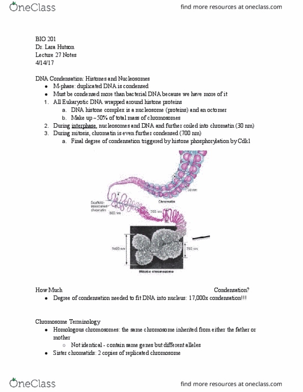
Heterochromatin: dark regions, contains more condensed material. Condensation of metaphase chromosomes results from several orders of folding of 30-nm chromatin fibers. Chromosomes that become visible during metaphase are duplicated structures; each metaphase chromosome consists of 2 sister chromatids, which are linked at centromere (constricted region) telomeres: end of chromatid. Centromere involved in mitosis: specific protein complexes build kinetochore, which recruits microtubules; microtubules separate chromatin in mitosis. Karyotype: the number, size, + shapes of the metaphase chromosomes, distinctive for each species. In metaphase, chromosomes distinguished by banding patterns + chromosome painting. Certain dyes selectively stain some regions of metaphase chromosomes more intensely, producing characteristic banding patterns, specific for individual chromosomes. G bands: produced when metaphase chromosomes are subjected briefly to mild heat or proteolysis, then stained with giemsa reagent (permanent dna dye: g-bands correspond to unusually low g+c content; creates a very reproducible pattern. Chromosome painting + dna sequencing reveal evolution of chromosomes.