PATH 3000 Lecture Notes - Lecture 17: Hepatic Veins, Portal Vein, Common Hepatic Artery

9 Mar 2020
School
Department
Course
Professor
Document Summary
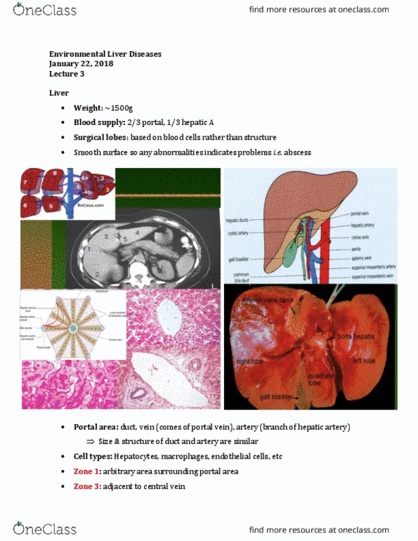
Path 3000 lecture 17 february 26, 2020 pancreas and liver pathology. Liver: describe liver anatomy (macro and microstructural) and function, discuss etiologies (viral, steatohepatitis, autoimmune, biliary, hereditary) of chronic liver disease. List clinical signs/symptoms of advanced chronic liver disease (cirrhosis) Identify the 2 most common primary liver malignancies. Pancreas: describe macro- and microanatomy of pancreas, distinguish function of exocrine vs endocrine pancreas, explain pathogenesis of diabetes. The liver weighs about 1. 45 kg, sits in the upper quadrant abdomen. There is a right and left lobe each separated into different segments by the hepatic venous drainage. The gallbladder is found in the posterior side. There are 3 major hepatic veins, left, middle, right, and drain into the ivc. Hepatocytes are big and pink because they have large cytoplasm and collagen. The lobule (portal central) is repeated over in the organ. A little bit of collagen acts as support. Some inflammatory cells are constantly present to monitor the state of the gut.