IMED3004 Lecture Notes - Lecture 10: Petrous Part Of The Temporal Bone, Neuroglia, Amenorrhoea

1 Nov 2020
School
Department
Course
Professor
Document Summary
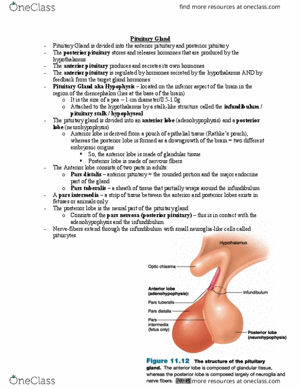
Anatomy: normally approx 1 cm (~ 500 mg) Situated in sella turcica of the sphenoid bone surrounded by dura, the sellar diaphragm" is a fold of dura that makes up the roof of the sella turcica) Lateral - cavernous sinus, internal carotid artery and cranial nerves iii, iv, v, vi. Superior - optic chiasm, hypothalamus, and third ventricle. Blood supply from superior and inferior hypophysial arteries, via portal vessels arising in capillaries of hypothalamus and posterior pituitary (therefore vulnerable to ischaemia) Anterior lobe (adenohypophysis) derived from rathke"s pouch in the pharynx 80%: an epithelial structure. Posterior lobe (neurohypophysis) derived from outpouching from the floor of the third ventricle (connection persists as pituitary stalk) 20%. Important because posterior pituitary has a direct anatomical connection to the brain and is made up of neural tissue. Histology- anterior pituitary: nests, cords and small acini of secretory epithelial (neuroendocrine) cells. Histology- posterior pituitary: modified glial cells (pituicytes) and nerve fibres extending from hypothalamus.