91500 Lecture Notes - Lecture 1: Renal Calyx, Sine Wave, Soft Palate

Document Summary
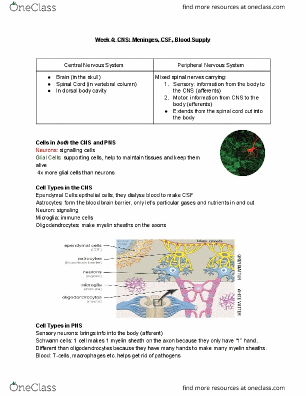
Dura (dense fibrous ct), pia (few cells thick, not largely noticeable, covers every tiny cleft) and arachnoid mater (loose, highly vascular, dips into clefts, most noticeable of the meninges) Oligodendrocytes small and round w/ clear halo surrounding. White matter ascending or descending from brain. Ependymal cells look like simple columnar epithelium. White spaces around nuclei = axoplasm of individual neurons. Surrounding nerve is ct layer of epineurium. Epithelium + loose ct under it are the tracheal mucosa. Submucosa underneath contains many seromucous glands and blood vessels. End of rings = bands of smooth trachealis muscle to adjust diameter of trachea. Outermost = ct & muscle comprise the adventitia. Bronchi > bronchioles > resp bronchioles > alveolar ducts > alveoli. Air space that is a common vestibule for several alveoli = alveolar duct. Red regions in alveolar wall = capillaries. Type 1 = small, spindly and comprises capillary wall. Type 2 = foamy contain surfactant (look like mesothelial cells)