BMED2402 Lecture 1: BMED2402 Full Lecture Notes

3 May 2018
School
Department
Course
Professor

BMED2402 Alicia Ngoc Diep Ha
Lecture 1 – Histology of the nervous system
Outcome
Key Points
State what
neurohistology is.
Histology of the nervous system
State what a neuron is
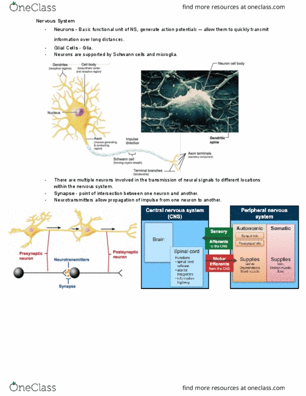
Neuron: cells found in NS, form interconnected network.
• Sense external (somatic) and internal (visceral) environments
• Process info and act on environments
• 3 ways of describing them – morphological, location, connection networks
Define the histological
spaces within the brain
• Function: mediate passage of mobile cells and molecules between spaces
• interstitial/ extracellular space: between neuronal and glial cells
• intravascular space: blood vessels
• intraventricular space: within ventricles
• subarachnoid space: between pia and arachnoid
• intracellular space: within cells in some neurons and glia
• Potential spaces: subdural and extradural space in relation to meninges
• Relative barriers: between spaces of mobile cells and molecules, formed by
capillary endothelium, glia limitans, ependyma and arachnoid
Discuss the various ways
in which cells of the
nervous system
including neurons are
characterized (classified,
described), give
examples.
Description and classification of cells in the NS
1. Function
2. General location – e.g. PNS, CNS
3. Specific location – e.g. layer of cerebral cortex
4. General connections – eg. Peripheral motor, sensory, central
intermediate/integrative
5. Polarity
6. Size
7. Shape of body or dendritic tree
8. Axon length
9. Transmitter
10. Electrical behavior
Give an overview of the
techniques
(microscopes, stains)
used
to visualize the cells of
the nervous system
find more resources at oneclass.com
find more resources at oneclass.com

BMED2402 Alicia Ngoc Diep Ha
Discuss and draw the
morphological
characteristics (internal
and external levels) of a
typical neuron including
the synaptic r
egion and correlate the
structure(s) with
function
• Has nucleus, nucleolus, ER, ribosomes, mitochondria, Golgi, lysosomes,
structural proteins, filaments, tubules and secretory vesicles
• Euchromatic nucleus (less active)
• Prominent nucleus (rRNA)
• Extensive ER – Nissl substance
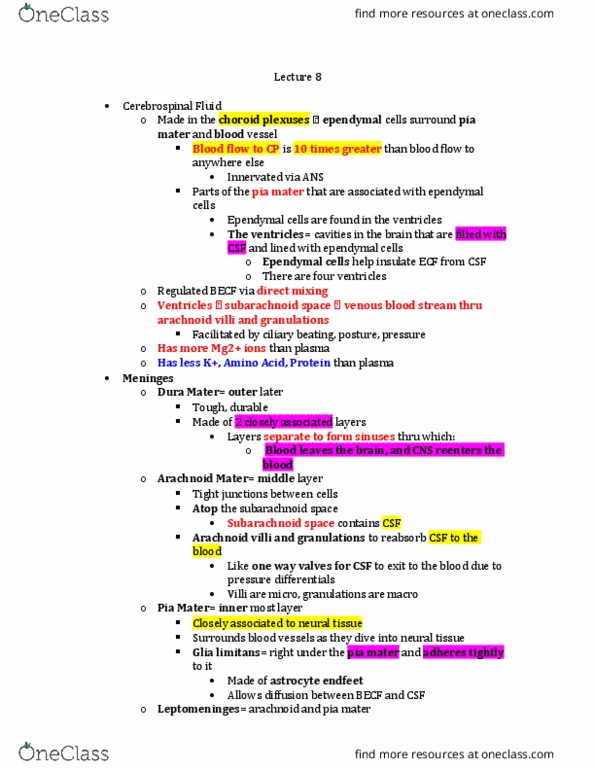
• Synapse – region for chemical transmission
Discuss and give
examples of the
variation in the structure
and
function of neurons
Variation in external appearance
• Axonal length
- Short for interneurons (Golgi type 2) within gray matter
- Long for projection (Golgi type 1) between gray matter
Discuss in principle the
connections of neurons
to one another
both locally
(microcircuits) and at a
distance (macrocircuits)
• Microcircuit (cytoarchitecture) – within gray matter
• Macrocircuit (part of functional system) – between gray matter
Give examples of the
histological organization
of neurons in
gray and white matter
eg the cerebral and
cerebellar cortex,
spinal cord gray matter,
and the retina
Cerebellar cortex
• Folded sheet of 3 layers
- Inner granular layer granule cell (interneuron)
- Intermediate Purkinje layer (projection neuron)
- Outer molecular layer – mainly cell processes
• Other types of interneurons – glial cells
Cerebral cortex
• Sheet of gray matter, mostly 6 layers
1. Molecular layer (most superficial)
2. External granular layer
3. External pyramidal layer
4. Internal granular layer
5. Internal pyramidal layer
6. Polymorphic/multiform layer (deepest)
Spinal cord gray matter
• H of gray matter in centre of SC
find more resources at oneclass.com
find more resources at oneclass.com

BMED2402 Alicia Ngoc Diep Ha
• LM appears relatively uniform meshwork of neurons, glia and capillaries
• Ventral horn – contains large cell bodies of multipolar lower (alpha) motor
neurons innervate skeletal muscle
Retina
• Forms inner incomplete layer of eye
• Other 2 layers – choroid and sclera/cornea
• Microscopically composed of 10 layers
• Direct pathway – photoreceptors (cones and rods – have special transducers
part of PNS), bipolar neurons and ganglion cell neurons
• Modulating interneurons, horizontal and amacrine cells
• Special type of glia/supporting cell – Muller cell
Discuss and draw the
morphological
characteristics of the
supporting cells
(astrocytes,
oligodendrocytes,
microglia,
ependyma, schwann
cells, satellite cells,
perineural cells) and
other cells (pia,
arachnoid, dura,
capillaries) of the
nervous
system and correlate
structure and location
with function
Astrocytes
• In CNS
• Two types – fibrous (in white matter) & protoplasmic (in gray matter)
• Medium – small spherical cells, many short processes, end feet
• Feed adjacent to capillaries – help mediate BBB
• Function: regulation of ion and transmitter
content in interstitial space, maintenance of
BBB and formation of scar tissue
Oligodendrocytes
• In CNS
• Mainly in white matter, near proximal end of
projection neuron axons
• Processes wrap around axons to form myelin sheaths
• Single oligodendrocytes can myelinate several axons
Microglia
• In CNS, small cells with elongated nuclei
• Derive from mononuclear cells
• Only representative of immune system in brain
• Macrophages – can leave and enter bloodstream
Ependymal cells
• In CNS
• Form layer of cuboidal cells, line ventricles and spinal canal of CNS
• Have villi and cilia on luminal surfaces
• Function unknown
Schwann cells
• In PNS
• Elongated cells wrap around segment of one axon (myelinated) or multiple
axon invaginated (unmyelinated)
• Increases rate of electronic impulse conduction – limits leakage of charge
from axonal membrane
Satellite cells
• In PNS
• Small cells, surround neuronal cell bodies in somatic
sensory and autonomic ganglia
find more resources at oneclass.com
find more resources at oneclass.com
Document Summary
Lecture 1 histology of the nervous system. Discuss the various ways in which cells of the nervous system including neurons are characterized (classified, described), give examples. Give an overview of the techniques (microscopes, stains) used to visualize the cells of the nervous system. Discuss and draw the morphological characteristics (internal and external levels) of a typical neuron including the synaptic r egion and correlate the structure(s) with function. Discuss and give examples of the variation in the structure and function of neurons. Discuss in principle the connections of neurons to one another both locally (microcircuits) and at a distance (macrocircuits) Short for interneurons (golgi type 2) within gray matter. Long for projection (golgi type 1) between gray matter: microcircuit (cytoarchitecture) within gray matter, macrocircuit (part of functional system) between gray matter. Cerebellar cortex: folded sheet of 3 layers. Outer molecular layer mainly cell processes: other types of interneurons glial cells.