HUBS2103 Lecture Notes - Vertebral Artery, Carotid Canal, Ventral Anterior Nucleus

1 Jul 2018
School
Department
Course
Professor

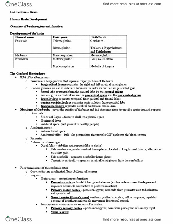
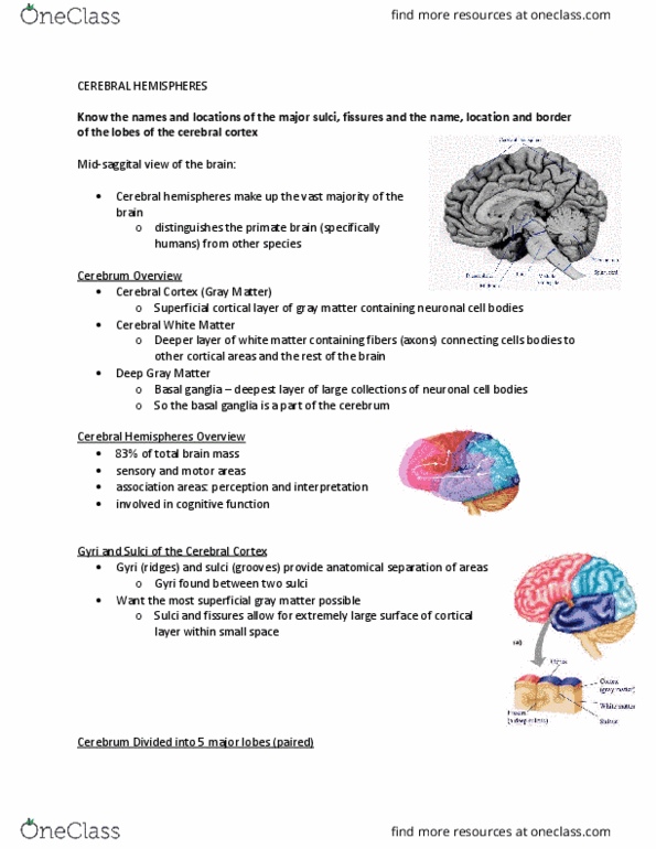
Cerebral Cortex & Meninges
Cerebral Cortex
• Most superficial layer of cerebrum
• Made up of grey matter
• Not to be confused with cerebellar cortex (superficial layer of cerebellum)
• 80% brain's total mass
• Gyri (multiple gyrus') are the raised areas and the sulci (multiple sulcus') are the indents
• Larger grooves are called fissures which divide the cerebral cortex into lobes
• The medial longitudinal fissure divides the cortex into left and right hemispheres
1. Motor cortex
• Primary motor cortex found in pre-central gyrus
o Main contributor to conducting neural impulses for the execution of movement
o Neurons that travel FROM this region will either go:
• Cortico-spinal tract
▪ To spinal cord for movements of body
• Cortico-bulbar tract
▪ To brainstem for movements of head, neck and face
• Non-primary motor cortex
o Divided into two regions:
• Supplementary motor cortex
▪ Sequence and selection of movement
• Pre-motor cortex
▪ PLANNING rather than execution of movement
find more resources at oneclass.com
find more resources at oneclass.com

find more resources at oneclass.com
find more resources at oneclass.com

2. Association cortex
• Posterior parietal (sometimes referred to as in motor cortex)
o Transforming sensory information into commands
o Motor planning
3. Somatosensory cortex
• Primary somatosensory cortex
o Post-central gyrus
o Processing somatic sensations:
• Touch
• Proprioception
• Nociception (pain)
• Temperature
o When body senses one of these, information is first sent to THALAUMS, then primary
somatosensory cortex
o Divided into four sections
• 3a
▪ Majority of somatosensory input from thalamus & does initial
processing
▪ Proprioceptors
• 3b
▪ Majority of somatosensory input from thalamus & does initial
processing
▪ Touch sensations
▪ Sends info to areas 1/2
• 1
▪ More complex processing
• 2
▪ More complex processing
▪ Proprioception
4. Visual Cortex
• located in occipital lobe
• Visual information goes through:
1. Eye
2. Lateral geniculate nucleus (in thalamus)
3. Visual cortex
• Primary visual cortex (V1)
o receives sensory input from thalamus
• The extrastriate areas
o V2
o V3
o V4
o V5
find more resources at oneclass.com
find more resources at oneclass.com
Document Summary
Not to be confused with cerebellar cortex (superficial layer of cerebellum) Gyri (multiple gyrus") are the raised areas and the sulci (multiple sulcus") are the indents. Larger grooves are called fissures which divide the cerebral cortex into lobes. The medial longitudinal fissure divides the cortex into left and right hemispheres: motor cortex. Main contributor to conducting neural impulses for the execution of movement. Neurons that travel from this region will either go: To brainstem for movements of head, neck and face. Planning rather than execution of movement: association cortex. Posterior parietal (sometimes referred to as in motor cortex) When body senses one of these, information is first sent to thalaums, then primary somatosensory cortex. Majority of somatosensory input from thalamus & does initial processing. Proprioception: visual cortex located in occipital lobe. Located on superior temporal gyrus in temporal lobe. Point-to-point input from ventral/ anterior division of the medial geniculate complex.