MEDI7111 Lecture Notes - Focal Segmental Glomerulosclerosis, Peripheral Edema, Iga Nephropathy


Renal 2
Pathology of the Kidney and Urinary Tract
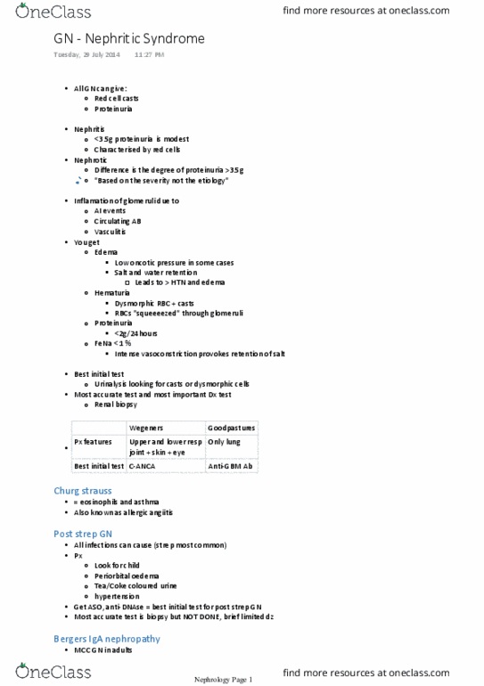
Nephritic Syndrome
Nephrotic Syndrome
Haematuria
- blood may be microscopic or
macroscopic
- Red cell casts: distinguishing feature,
form in nephrons and indicate
glomerular damage
- Dysmorphic red cells → indicates
glomerulus as the site of bleeding
- Podocytes develop large pores which
allow blood and protein through
Some proteinuria
Mild Hypertension
Azotaemia
- Abnormally high blood urea & nitrogen
(BUN)
- Leads to decreased blood flow,
decreased in GFR, stimulation of RAS
Oliguria
Lesions causing nephritic syndrome all have
increased cellularity within the glomeruli,
accompanied by a leukocyte infiltrate.
This causes inflammation which injures capillary
walls which let red blood cells leak into urine.
This hemodynamic change causes decreased GFR
and stimulation of RAS.
Proteinuria (> 3.5g in 24hrs)
Hypoalbuminaemia
- Albumin is lost in urine
- Binds cations, water, hormones, fatty
acids, and others to regulate the osmotic
pressure of blood
- Gaps between podocytes allow proteins
to escape
Oedema
- Due to loss of albumin (Intravascular
oncotic pressure decreased → Fluid leaks
out of vessels)
- Ascites, non-pitting peripheral oedema,
periorbital oedema
Hyperlipidaemia
- Due to loss of albumin, liver has to
compensate for lower products in the
blood and uses lipids to ‘bulk’ it out
- Fatty changes in the liver are also present
Nephrotic is often more dangerous than
nephritic.
Conditions presenting with Nephritic Syndrome
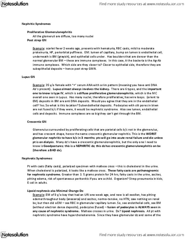
Post streptococcal GN
IgA Nephropathy
Rapidly progressive/ crescentic GN
Membranoproliferative GN
Goodpasture’s Syndrome
Vasculitis disorders – Wegner’s Granulomatosis,
Microscopic Polyangiitis, Churgg-Strauss disease,
Henoch-Schönlein purpura
Conditions presenting with Nephrotic Syndrome
Primary causes
Minimal change Glomerulonephritis
Focal Segmental Glomerulosclerosis
Membranous Glomerulonephritis.
Secondary causes
SLE, Hep B & C, HIV
Diabetes Mellitus
Malignancy (particularly CML)
Kidney Disease
Acute vs Chronic Kidney Disease
Acute kidney disease generally effects a single anatomical compartment (e.g. glomerulus, tubules,
interstitium, vasculature etc.) of the kidney, resulting in a loss of a specific function that leads to a
recognisable clinical presentation. Acute kidney injury can occur as part of a local disease process or
secondary to the failure of other organ systems (e.g. MODS). Compensation for the disease occurs to
limit dysfunction.
Chronic renal disease results from failure of the acute compensatory mechanisms to maintain
adequate function in the injured kidney. This can cause further damage to the kidney, for example,
damage to the glomerulus causing ischemic loss of downstream tubules leading to necrosis and
inflammation – further limiting the functionality of the kidney. Alternately, obstruction of urinary
outflow can cause back pressure in the filtration units leading to glomerular damage. Chronic
damage to the filtration units results in a loss of renal function as these units are unable to be
regenerated. Once GFR reaches 30-50% of normal, progression to renal failure is inevitable.
Clinical Presentation of Glomerular Disease
There are four primary presentation patterns for glomerular disease:
• Nephrotic syndrome
o Massive proteinuria (>3.5g/day)
o Hypoalbuminaemia (<3g/dL)
o Generalised oedema
o Hyperlipidaemia
o Lipiduria
• Nephritic syndrome
o Acute onset macroscopic haematuria
o Mild to moderate proteinuria
o Hypertension
• Asymptomatic proteinuria/haematuria
• Rapidly progressing acute renal failure
o Acute nephritis
o Acute renal failure
o Proteinuria
When diagnosing a glomerular disease, a renal biopsy is very helpful in determining the cause. This
provide information as to the prognosis and treatment options for the patient, however many
glomerular diseases have the same morphology or overlapping morphologies with other glomerular
diseases. This is where clinical signs and symptoms help to distinguish disease types – something
that you leave to the nephrologists.

Nephritic Syndromes
Post Infectious GN
Epidemiology
• Most common in children 6-10 years (but does occur at other ages
too)
Risk Factors
• Streptococcal infection
o Appears 1-4 weeks post infection
o Only certain strains of Group A β haemolytic strep are
nephritogenic
Pathophysiology
• Global and diffuse pattern of disease
Clinical Features
• In Children: abrupt development of:
o Malaise
o Fever
o Nausea
o Haematuria (dysmorphic red cells/casts)
o Oliguria
o Hematuria (smoky/coke coloured urine)
o Mild to moderate hypertension
• In Adults:
o Sudden onset hypertension/oedema
o Elevated BUN (blood urea nitrogen)
Prognosis
• Children
o 95% make a full recovery
o <1% do not improve and progress to RPGN
o Remaining patients have a slow progression to chronic GN
• Adults
o 60% make a full recovery quite quickly
o Remainder have a less benign course:
▪ Persistent haematuria and hypertension
▪ Slow progression to chronic GN
▪ RPGN
Light Microscopy
Immunofluorescence
Electron Microscopy
Enlarged, hypercellular
glomeruli
• Infiltration of
leukocytes
• Proliferation of
Mesangial and
endothelial cells
Crescent formation in severe
cases
Red cell casts
Granular deposits of IgG & C3
Focal and sparse
“Humps” in the basement
membrane from immune
complex deposition
Will eventually fall off on their
own so no treatment is required
to cause resolution (self-limited
disease)
Document Summary
Red cell casts: distinguishing feature, form in nephrons and indicate glomerular damage. Dysmorphic red cells indicates glomerulus as the site of bleeding. Binds cations, water, hormones, fatty acids, and others to regulate the osmotic pressure of blood. Podocytes develop large pores which to escape allow blood and protein through. Abnormally high blood urea & nitrogen (bun) Leads to decreased blood flow, decreased in gfr, stimulation of ras. Lesions causing nephritic syndrome all have increased cellularity within the glomeruli, accompanied by a leukocyte infiltrate. This causes inflammation which injures capillary walls which let red blood cells leak into urine. This hemodynamic change causes decreased gfr and stimulation of ras. Due to loss of albumin (intravascular oncotic pressure decreased fluid leaks out of vessels) Due to loss of albumin, liver has to compensate for lower products in the blood and uses lipids to bulk" it out. Fatty changes in the liver are also present.