DEV2011 Lecture Notes - Lecture 10: Yolk Sac, Syncytiotrophoblast, Placenta

25 May 2018
School
Department
Course
Professor

Lecture 10 – Placenta and Extraembryonic Membranes
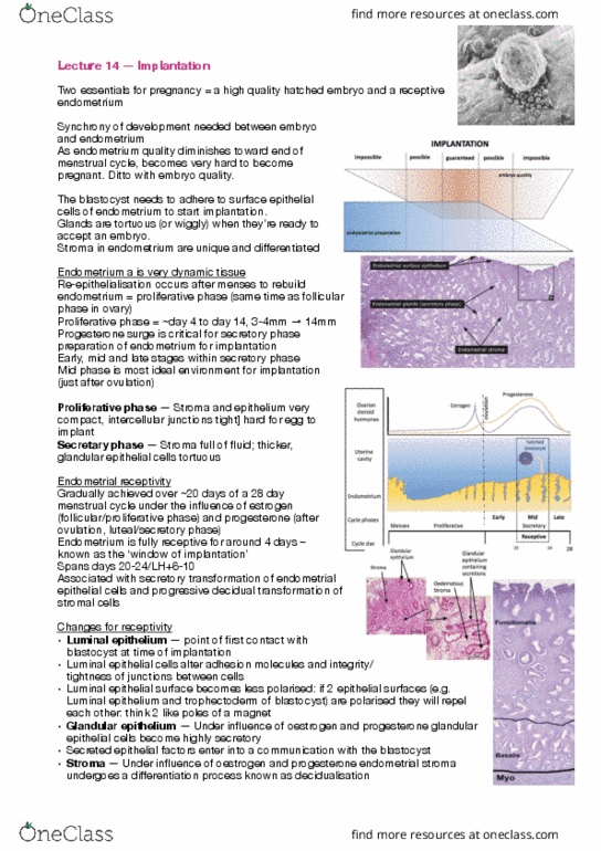
Uterine Receptivity
• Menstrual cycle is under control of ovarian hormone cycle
• Day 1: menses: drop of progesterone (sheds endometrium)
o Uterus undergoes proliferation ot rebuild
• Following ovulation: oestrogen is high → uterus undergoes secretion
o Glands secretes factors that promote attachment of blastocyst to uterus
• Uterus is only receptive for 4 days (days 6-10) during menstrual cycle
• If blastocysts enter uterus before or after receptive time frame, pregnancy
wont be established due to lumen epithelium wont be receptive to attachment
Implantation
• Blastocyst apposes then adheres to uterine luminal epithelium
o Trophoblast begins to invade between epithelial cells
• Decidual cell controls how far blastocyst invade
Cell Lineages
• Blue: inner cell mass
o Embryo proper
o Amnion
• Yellow: endoderm
o Yolk sac
• Red: mesoderm
• Green: trophectoderm
o Cytrotrophoblast
o Invasive trophoblast
o Syncytiotrophoblast
Amnion
• Surrounds fetus
• Liquid filled
o Cavity expands during gestation
o 1L ~33-34 weeks
• Amniotic membrane
o Composed of extraembryonic ectodermal cells lined with
nonvascualised extraembryonic mesoderm
• Role
o Protective buffer against mechanical injury
Yolk Sac
• Lined by extraembryonic endoderm
• Outside well vascularized extraembryonic mesoderm
• Birds and reptiles
o Provides yolk for nutrition
• Mammals
o Primordial germ cells arise in extraembryonic mesoderm near base of
allantois (3rd week)
o Become visible in lining of yolk sac → migrate to gonads
find more resources at oneclass.com
find more resources at oneclass.com

Allantois
• Endodermally lined ventral outpocketing of hindgut
• Other vertebrates: major respiratory organ and repository for urinary wastes
• Humans – vestige
o Respiration: blood vessels differentiated from mesodermal wall of
allantois
Human Placenta
• Haemochorial
o Villi and outer surface of chorionic plate bathed in maternal blood
• Fetus connected to placenta via umbilical cord
• Placenta invasive to uterine tissue
• Hematopoietic tissue
Cell Linages – Trophoblast
1. Synctritrophoblast
• Outer layer line vilous
• Multinucleated
• Bathed in maternal blood
2. Cytotrophoblast
• Beneath synctriotrophoblast
• Located in cell column – proliferative
• Invasive
o Extravillous trophoblast (EVT)
▪ Interstitial (iEVT)
▪ Endovascular (eVET)
Placenta Villi
• Villi floating in intervillous space
o Bathed in maternal blood
o Outer surface of villi – synctiotrophoblast
Anchorage to Decidua
• Anchors via synctiotrophoblast
• Placenta tries to create own blood
supply
• Invades and engulfing blood vessels
Mature Placenta
• Foetal circulation: deoxygeneated blood
Arterial Remodelling
• If you don’t remodel arteries properly
then placenta is compromised → diseases
• Volume hasn’t changed but pressure alters
Term Placenta
• Born 30 mins after fetus
• Maternal surface
find more resources at oneclass.com
find more resources at oneclass.com
Document Summary
If blastocysts enter uterus before or after receptive time frame, pregnancy wont be established due to lumen epithelium wont be receptive to attachment. Implantation: blastocyst apposes then adheres to uterine luminal epithelium, trophoblast begins to invade between epithelial cells, decidual cell controls how far blastocyst invade. Cell lineages: blue: inner cell mass, embryo proper, amnion, yellow: endoderm, yolk sac, red: mesoderm, green: trophectoderm, cytrotrophoblast, invasive trophoblast, syncytiotrophoblast. Amnion: surrounds fetus, liquid filled, cavity expands during gestation, 1l ~33-34 weeks, amniotic membrane, composed of extraembryonic ectodermal cells lined with nonvascualised extraembryonic mesoderm, role, protective buffer against mechanical injury. Allantois: endodermally lined ventral outpocketing of hindgut, other vertebrates: major respiratory organ and repository for urinary wastes, humans vestige, respiration: blood vessels differentiated from mesodermal wall of allantois. Human placenta: haemochorial, villi and outer surface of chorionic plate bathed in maternal blood, fetus connected to placenta via umbilical cord, placenta invasive to uterine tissue, hematopoietic tissue.