BMS1052 Lecture Notes - Lecture 12: Stapedius Muscle, Grey Matter, Eustachian Tube

30 May 2018
School
Department
Course
Professor

Week 5. Vision and the Eye II, Audition & the control of
movement
VISION AND THE EYE II
• Signals are passed to bipolar cells
• Horizontal cells also provide horizontal connection between photoreceptors and bipolar cells
• Photoreceptors are unusual as they hyperpolarise when receive preferred stimulus
• Receptive fields and bipolar cells
RF= egio of spae i hih light hages affet a ell’s MP
Bipolar cells have an antagonistic, centre surround RF structure
-in the dark, photoreceptors are depolarised and tonically active, increased light hyperpolarises
them (reduces output)
i.e. light off = more glutamate, light on = less glutamate
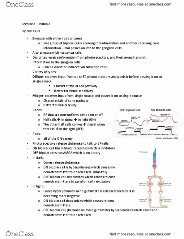
OFF bipolar cells
ON bipolar cells
o Hyperpolarised by light in RF centre
o Preserve sign of photoreceptors
- Glutamate from photoreceptors is
excitatory (depolarises cells)
-increased illumination = less glutamate ->
hyperpolarises cell
o Depolarised by light in RF surround
o Depolarised by light in RF centre
o Invert sign of photoreceptors
-glutamate from photoreceptors is inhibitory
(hyperpolarises)
-increased illumination = less glutamate ->
depolarises cell
o Hyperpolarised by light in RF
-antagonistic = centre and surround do the opposite thing
find more resources at oneclass.com
find more resources at oneclass.com

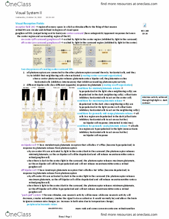
o bipolar cell receives direct inputs from blue photoreceptors -> contribute to RF centre
(directly activate bipolar cell)
o horizontal cell provides lateral inhibition between adjacent photoreceptors -> red
photoreceptors can indirectly inhibit bipolar cell
-get centre-surround inhibition
o Together, the red and blue photoreceptors form the neuronal receptive field – they are the
inputs.
o We have a center, and a surround, and light outside this center-surround receptive field
does ot affet the ipola ell’s fiig rate.
o Only horizontal cells are directly connected to bipolar cells -> account for RF centre
• Why is the center-surround structure important?
o Fist, it’s uiuitous i sesoy systes – improves spatial localisation ability.
-> helps locate and identify stimuli
o Moe ipotatly, it’s the uildig lok of a oe sophistiated isual syste.
• Cell types:
Photoreceptors
o Light transduction via G-protein coupled
opsin molecule
o Depolarised in darkness
o Non spiking -> glutamate release decreases
as light levels increase
o No rods in fovea
o Fewer cones in periphery
Bipolar cells
o OFF-bipolar: hyperpolarised by light on RF
centre
o ON-bipolar: depolarised by light on RF centre
o Non-spiking -> graded membrane potentials
Horizontal cells
o Bidirectional
o Inhibitory connections to photoreceptors
o Mediates bipolar cell RF surrounds
Retinal ganglion cells
o Centre-surround RF
o Spiking responses
o Retinal output cells
o Convergence ratio of inputs from
photoreceptors affects visual acuity
find more resources at oneclass.com
find more resources at oneclass.com

• Information flow: photoreceptor -> bipolar cells -> ganglion cells
• Centre-surround RF can mostly explain the illusions:
o Lateral inhibition
->fundamental mechanism in all sensory systems
-> improves spatial localisation
->improves stimulus identification
o Relative illumination is important – both spatially and temporally
->increase and decrease in light levels are significant
• Functional segregation: retinal ganglion cells project to many places
o *Lateral geniculate nucleus (LGN): gateway to cortex and conscious vision
-90% of retinal projection
o Pretectum: reflexive eye movements and pupil size
o Superior colliculus: controls eye and head orienting responses
o Suprachiasmatic nucleus: in hypothalamus, circadian rhythms
• Field of view vs hemifield:
Visual field
Hemifield
o The image on the right (nasal) portion of the
left eye projects to the right side of the brain
o the image on the right (temporal) portion of
the right eye, projects to the right side of the
brain (eg. The left visual cortex represents
the right visual field.)
o Signals from each visual hemifield
target the contralateral LGN and primary
visual cortex
o left or right of the vertical meridian.
find more resources at oneclass.com
find more resources at oneclass.com
Document Summary
Vision and the eye ii, audition & the control of movement. Vision and the eye ii: signals are passed to bipolar cells, horizontal cells also provide horizontal connection between photoreceptors and bipolar cells, photoreceptors are unusual as they hyperpolarise when receive preferred stimulus, receptive fields and bipolar cells. Rf= (cid:396)egio(cid:374) of spa(cid:272)e i(cid:374) (cid:449)hi(cid:272)h light (cid:272)ha(cid:374)ges affe(cid:272)t a (cid:272)ell"s mp. Bipolar cells have an antagonistic, centre surround rf structure. In the dark, photoreceptors are depolarised and tonically active, increased light hyperpolarises them (reduces output) i. e. light off = more glutamate, light on = less glutamate. Off bipolar cells: hyperpolarised by light in rf centre, preserve sign of photoreceptors. Glutamate from photoreceptors is excitatory (depolarises cells) Increased illumination = less glutamate -> hyperpolarises cell. On bipolar cells: depolarised by light in rf centre. Increased illumination = less glutamate -> depolarises cell: depolarised by light in rf surround, hyperpolarised by light in rf.