PSYC10003 Lecture Notes - Lecture 9: Nociceptor, Capsaicin, Bulbous Corpuscle

6 Jun 2018
School
Department
Course
Professor

PSYC10003 – MIND, BRAIN, & BEHAVIOUR 1
BEHAVIOURAL NEUROSCIENCE
Lecture 9 (Week 3 . 3): The Auditory & Somatosensory Systems
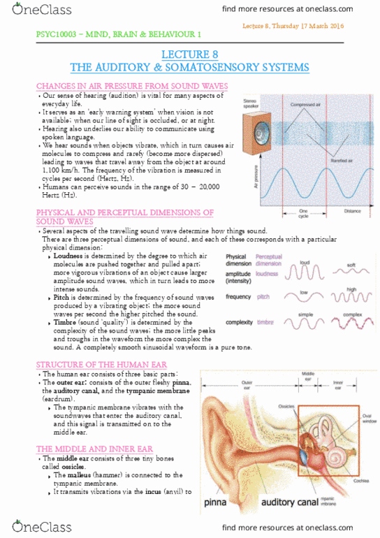
Sound waves: we hear sounds when objects vibrate, causing air molecules to compress
& rarefy (become more dispersed) producing waves that travel away from the object
• Frequency of vibration is measured in cycles per second (Hertz, Hz)
• Perceptual dimensions / aspects of sound:
• Loudness: degree to which air molecules are pushed together & pulled apart; more
vigorous vibrations of object, larger amplitude sound waves, more intense sounds
• Pitch: the frequency of sound waves produced by a vibrating object; the more sound
waves per second the higher pitched the sound
• Timber: (sound ‘quality’) the complexity of the sound waves; the more little peaks & troughs in
the waveform the more complex the sound. Pure tone: a
completely smooth sinusoidal waveform
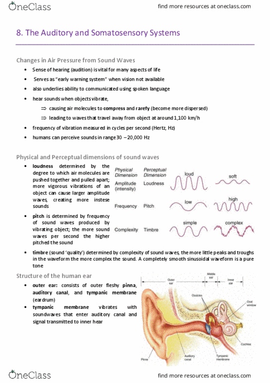
Human Ear:
• Outer ear: consists of outer fleshy pinna, auditory canal,
& tympanic membrane (eardrum). Tympanic membrane
vibrates with the soundwaves that enter the auditory canal,
& this signal is transmitted to the middle ear
• Middle ear: consists of 3 tiny bones (ossicles): malleus is connected to the tympanic
membrane, & transmits vibrations via the incus to the stapes, which is connected to the cochlea
• Inner ear: consists of the cochlea, bony structure with 2 small membranes that form windows on its
fluid filled interior, & contains receptors for analysing sounds. Stapes connects to the oval window,
& sound waves cause the stapes to move in & out, moving the fluid over receptors
inside the cochlea. The round window (2nd window) allows the fluid to move
Basilar Membrane: spiralled sheet of tissue, containing auditory receptors. It sits in the
centre of the cochlea, & runs from its base to its apex (diagram to right)
Organ of Corti: runs length of cochlea (diagram →) composed of basilar membrane,
receptors (hair cells), & rigid shelf over top (tectorial membrane)
• Hair cells: movement of basilar membrane
toward tectorial membrane bends stereocilia
(cilia on hair cells) through direct contact with
tectorial membrane or fluid motion, resulting in
receptor potentials - converting sound waves →neural signals
Spiral ganglion: stimulated by neurotransmitters from the hair cells.
Coded frequency of basilar membrane: different
frequencies = different places. Diff. spiral ganglions
code particular frequencies along the membrane
Pathway to Auditory Cortex: auditory nerve
transmits from cochlea to brainstem (Medulla).
• The neural info undergoes several stages of processing before reaching the
primary auditory cortex
• Info from each ear goes to both hemispheres – essential for localising sounds
find more resources at oneclass.com
find more resources at oneclass.com
Document Summary
Sound waves: we hear sounds when objects vibrate, causing air molecules to compress. Human ear: outer ear: consists of outer fleshy pinna, auditory canal, Tympanic membrane vibrates with the soundwaves that enter the auditory canal, & sound waves cause the stapes to move in & out, moving the fluid over receptors inside the cochlea. The round window (2nd window) allows the fluid to move. Basilar membrane: spiralled sheet of tissue, containing auditory receptors. It sits in the centre of the cochlea, & runs from its base to its apex (diagram to right) Spiral ganglion: stimulated by neurotransmitters from the hair cells. Coded frequency of basilar membrane: different frequencies = different places. Diff. spiral ganglions code particular frequencies along the membrane. Pathway to auditory cortex: auditory nerve transmits from cochlea to brainstem (medulla): the neural info undergoes several stages of processing before reaching the primary auditory cortex. Info from each ear goes to both hemispheres essential for localising sounds.