PSYC10003 Lecture Notes - Lecture 4: Dysarthria, Brainstem, Myasthenia Gravis

6 Jun 2018
School
Department
Course
Professor

PSYC10003 – MIND, BRAIN, & BEHAVIOUR 1
BEHAVIOURAL NEUROSCIENCE
Lecture 4 (Week 2 . 1): The Synapse
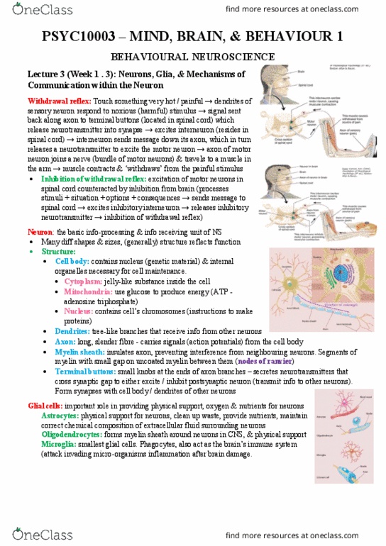
Synapse:
• Types:
• Axodendritic: terminal button synapses with a dendrite
• Axosomatic: terminal button synapses with the cell body (soma)
• Axoaxonic: terminal button synapses with the axon
• Structure:
• Presynaptic membrane: membrane of presynaptic terminal button
• Postsynaptic membrane: membrane of postsynaptic neuron
• Dendritic spine: ridge on dendrite of postsynaptic neuron, with
which terminal button from presynaptic neuron forms a synapse
• Synaptic cleft: tiny gap between pre & postsynaptic membrane
• Synaptic vesicles: tiny balloons filled with NT molecules; found in
terminal button release zone
• Microtubules: long tubes running down the axon that guide the transport of synaptic vesicles
from soma to the axon terminal
• Release zone: part of interior of presynaptic membrane that synaptic vesicles fuse with to
release their neurotransmitter into the synaptic cleft
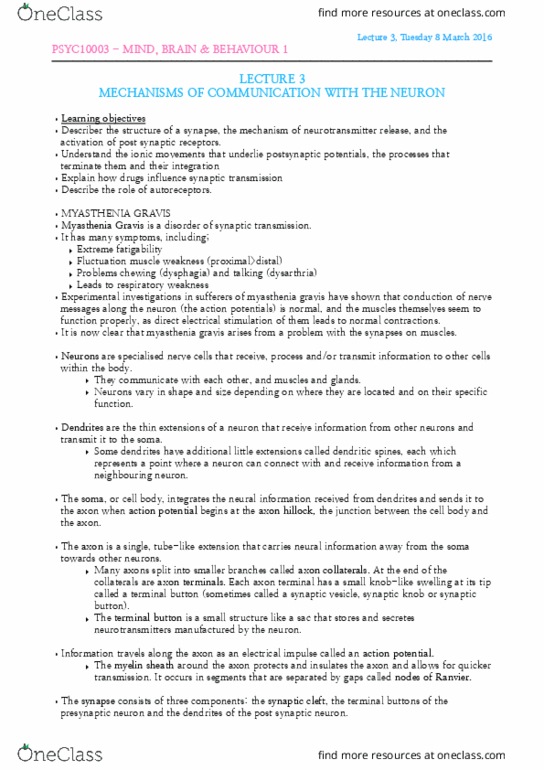
Synaptic transmission: when AP reaches terminal buttons, it triggers the release of a chemical message
(neurotransmitters). NTs diffuse across the synaptic cleft.
• NT has excitatory effect: depolarises the neuron, triggers action potential
• NT has inhibitory effect: hyperpolarises the neuron, doesn’t trigger an action potential
• Excitatory postsynaptic potentials (EPSPs): depolarise the postsynaptic cell
membrane, increasing likelihood that an AP will be triggered in postsynaptic neuron
• Inhibitory postsynaptic potentials (IPSP): hyperpolarise the postsynaptic cell
membrane, decreasing likelihood that an AP will be triggered in postsynaptic neuron
• Release of Neurotransmitter: AP causes synaptic vesicles to move towards release
zone (guided by protein structures that act as ropes). There’s an influx of calcium Ca2+
ions into presynaptic neuron, inducing fusion of synaptic vesicle & cell membrane. NT
molecules are released into synaptic cleft (diagrams to right)
• Receptor activation on post-synaptic neuron: NTs attach to specific binding
sites of postsynaptic receptors (located in the membrane of postsynaptic cell),
opening NT-dependent ion channels in the postsynaptic cell, permitting flow
of specific ions into / out of the postsynaptic neuron.
• *NTs open ion channels either directly or indirectly*
• Direct method: involves receptors equipped with their own binding sites
(ionotropic receptors), which a NT molecule binds to open the channel
• Movement of ions: whether postsynaptic potential is excitatory or inhibitory is determined not by
the NT that’s released into the synapse, but by the specific ion channel that it opens
• Sodium (Na+) channels: important for triggering excitatory postsynaptic potentials
• Potassium (K+) channels: K+ ions leave the neuron (since there’s slight excess of K+ inside
cell during rest), hyperpolarising the cell (inhibitory postsynaptic potential)
• Chloride (Cl-): inflow of Cl- causes hyperpolarisation (inhibitory postsynaptic potential)
find more resources at oneclass.com
find more resources at oneclass.com