ANAT30007 Lecture Notes - Lecture 25: Sesamoid Bone, Subtalar Joint, Deltoid Ligament

25 Jul 2018
School
Department
Course
Professor
Document Summary
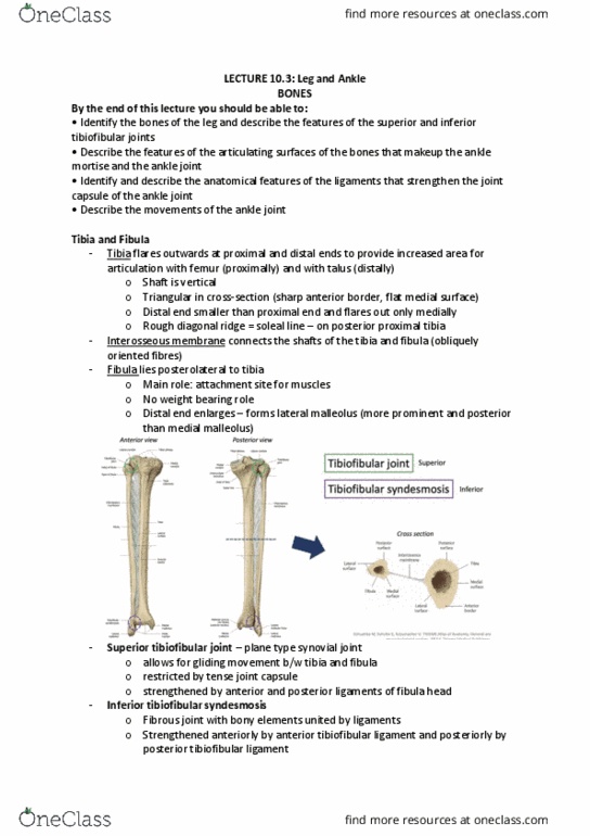
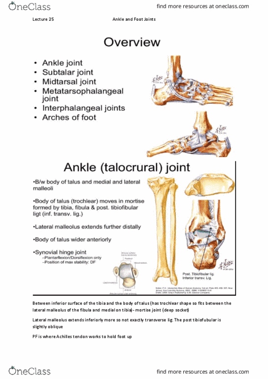
Between body of talus and medial and lateral malleoli of the tibia. Body of talus (trochlear) moves in deep socket (mortise) formed by tibia, fibula and posterior tibiofibular ligaments (inferior transverse ligament) Lateral malleolus extends further distally so the joint sits slightly oblique. Position of maximal stability = dorsiflexion because anterior talus is wider. Plantarflexion is supported by achilles tendon (picks up heel of foot) Capsule and synovial membrane attaches to articular margins and covers neck of talus anteriorly. Collateral ligaments (medial and lateral: both triangular (apex at malleoli, blend with joint capsule, facilitate df/pf movement. Mcl (deltoid ligament: very strong, four parts that merge together for continuous support, superficial (3 parts) with continuous attachment tibionavicular, tibiocalcaneal, posterior tibiotalar, deep(1 part) anterior tibiotalar, limits overeversion. Lcl (3 discrete parts: anterior talofibular (most commonly sprained, excessive inversion, calcaneofibular, posterior talofibular, commonly sprained because of discrete parts. Dorsiflexion = tibialis anterior, extensor digitorum longus, extensor hallucis longus.