ANAT30007 Lecture 10: [H1/91 notes] Principles of Imaging the Musculoskeletal System A

10 Jun 2018
School
Department
Course
Professor

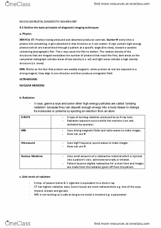
Speciality of medical imaging
•
Originally using Rotgen's "new" rays (X-rays 1895)
•
Radiographs thus radiology
•
No longer reliant on X-rays
○
Expanded dramatically over last 20-40 years to include many different modalities
•
Radiology
2 aspects to medical imaging
Structure: Anatomy
Function: How is that structure working? Increasingly important
Plain radiography
•
CT
•
US
•
MRI
•
Gamma imaging
○
PET
○
NM (nuclear medicine)
•
Modern medical imaging
Usually electromagnetic radiation
○
Acoustic
○
Energy source
•
Interaction of that energy with the body
•
Detection device for that energy
•
Image display
•
Basis of all medical imaging
Energy sources
4.1 Principles of Imaging the Musculoskeletal System
Monday, 23 March 2015
12:27 am
Locomotor Page 1

X-rays (radiographs, CT)
○
γrays (NM)
○
positrons (PET) decay →γrays
○
Ionising - detach electrons from atoms (thus can cause injury to body)
•
sound (US)
○
FM radio/magnetism (MRI)
○
non-ionising
•
Energy sources
Digital image
pixels
•
vacuum tube with electric current between a filament and target (cathode and anode),
current releases energy, forming X-rays
○
Crookes tube - X-rays
•
transmission of X-rays
○
e-density of tissues i.e.atomic number& []
•
more recently “x-ray detector”
○
Photo graphic film(negative)
•
more recently computer monitor
Light box
•
Plain radiography
Locomotor Page 2

more recently computer monitor
○
photoelectric interaction: atom is ionised; compton interaction: X-ray is deflected and
becomes noise
•
Bone: dense and contains Ca (high atomic number)
•
Soft tissue
•
Locomotor Page 3
Document Summary
Expanded dramatically over last 20-40 years to include many different modalities. Ionising - detach electrons from atoms (thus can cause injury to body) Rays (nm) positrons (pet) decay rays non-ionising sound (us) Crookes tube - x-rays vacuum tube with electric current between a filament and target (cathode and anode), current releases energy, forming x-rays e-density of tissues i. e. atomic number& [] transmission of x-rays. Locomotor page 2 more recently computer monitor photoelectric interaction: atom is ionised; compton interaction: x-ray is deflected and becomes noise. Bone: dense and contains ca (high atomic number) Right: lung replaced by consolidation/fluid denser than air. Locomotor page 4 energy source, multiple detectors - rotate around each slice. Crookes tube - x-rays e-density of tissues transmission of x-rays. X-ray detector solid-state scintillation crystal gas ionization chamber. Transmission of high frequency sounds acoustic differences between different tissues; sound is reflected back when there is a difference. Piezoelectric crystal - converts sounds to an electric signal.