ANAT30007 Lecture Notes - Lecture 18: Anatomical Terms Of Location, Interosseous Membrane, Metatarsal Bones

20 May 2018
School
Department
Course
Professor

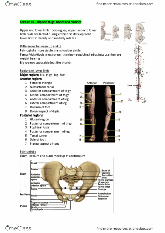
Leg
Lateral: foot evertors
•
Anterior: foot dorsiflexors
•
Posterior: foot plantar flexors
•
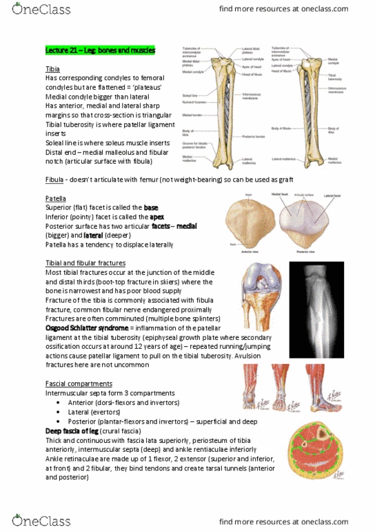
Tibia and fibula
7.3 Leg: Bones and Muscles
Monday, 20 April 2015
9:24 pm
Locomotor Page 1

Lateral/medial tubercles of intercondylar eminence: cruciate ligaments attach in front
and behind
○
Tibial plateau makes contact with femur
○
Condyles, soleal line (origin of soleus)
○
Sharp anterior (subcutaneous) and lateral borders
○
Tibial tuberosity: patellar ligament
○
Distal fibular notch
○
Medial and lateral malleoli form mortise of ankle joint
○
Tibia: robust bone that takes most of weight
•
Head contacts tibia
○
Interosseous membrane on medial border
○
Can be harvested for bone graft
○
Distal depression for tendons of evertors
○
Fibula: long and gracile, non-weight bearing
•
Patella
Locomotor Page 2
Document Summary
Tibia: robust bone that takes most of weight. Lateral/medial tubercles of intercondylar eminence: cruciate ligaments attach in front and behind. Medial and lateral malleoli form mortise of ankle joint. Lateral facet larger than medial - lateral condyle of femur projects more proximally to stop lateral dislocation. Most tibial fractures occur at junction of middle & inferior thirds (boot-top fracture in skiers from bending stress) - bone narrowest & poor blood supply. Fractures often comminuted (more than 2 separate bone components) Fracture of tibia commonly associated with fibula fracture. Common fibular nerve endangered proximally, supplies anterior leg. Osgood-schlatter disease: inflammation of the patellar ligament at the tibial tuberosity. 1 flexor (medial malleolus to calcaneus): holds plantar flexor tendons, digit flexors. 2 extensor: superior/superficial (across malleoli), inferior/deeper (y-shape, meets with evertor retinaculum) - dorsiflexion and extension of toes. Muscles surrounded by "tight" fascia - unable to expand on exertion. If swelling in neurovascular bundle, structures susceptible to compression - "compartment" syndrome.