HUMB1000 Lecture Notes - Lecture 2: Haematoxylin, Microtome, Eosin

3 Aug 2018
School
Department
Course
Professor
Document Summary
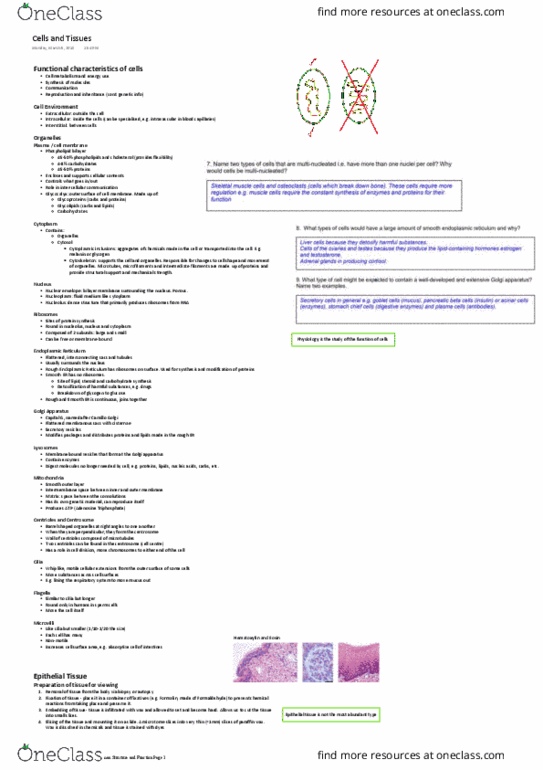
Haematoxylin and eosin is a stain that presents nuclei as purple and other structures pink. Points to consider: plane tissue has been cut-transverse, sagittal or frontal, the magnification of the tissue/image (higher=more description) Primary tissues (epithelial, connective, muscle, nervous: epithelial tissue/epithelium-covers. A alveoli of lungs, kidney glomeruli, serous membranes of. Simple cuboidal epithelium- single layer of cubed shaped cells (some have microvilli to aid absorption) Absorption, secretion(because the are fatter) and movement (due to flagella) pleura, pericardium and peritoneum. Simple columnar-shaped or tall cells with round to oval nuclei(some have cilia) Transitional epithelium- resembles stratified squamous and stratified cuboidal cells depending on its state. Stratified squamous-basal cells are cuboidal or columnar and become progressively flatter (squamous) as you move to the surface. Accommodate changes in fluid volume of the organs (depending on amount of fluid will depend on the state) Urinary bladder, ureter and upper part of urethra. Keratinized (water protection): sole of feet, palm of hands, skin.