HUMB1000 Lecture Notes - Lecture 6: Papillary Muscle, Interventricular Septum, Heart Rate

21 Jun 2018
School
Department
Course
Professor

C6 L1 – Anatomy of the Cardiovascular
System:
Functions:
- Transports fluids, nutrients, waste products, gases and hormones throughout the body
- Exchange materials between blood, cells and extracellular fluid
- Role in immune response, blood pressure and regulation of temperature
The Heart:
Functions:
- Generates blood pressure (moves blood through vessels via contractions
- Routing blood (separates pulmonary and systemic circulations)
- Ensuring one way blood flow
- Regulating blood supply
- Changes to match needs
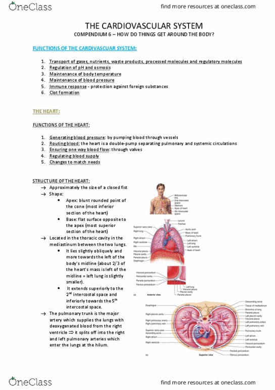
Location:
- Apex (most inferior part)
- Base (most superior, flat part)
- Located in thoracic cavity in mediastinum
- 2/3 on left side
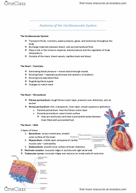
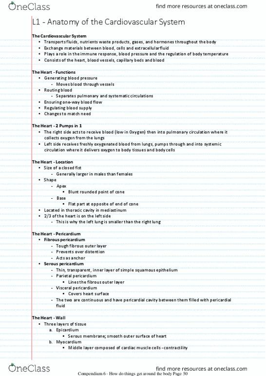
Pericardium:
- Double layered sac surrounding the heart
- Fibrous pericardium tough fibrous outer layer, prevents over distention, acts as an anchor
- Serous pericardium thin, transparent, inner layer, simple squamous epithelium
oParietal pericardium lines fibrous outer layer
oVisceral pericardium convers heart surface
oPericardial cavity filled with pericardial fluid
Wall:
- Epicardium serous membrane, smooth outer surface
- Myocardium middle layer composed of cardiac muscle cells, contractionary
- Endocardium smooth inner surface of heart chambers
- Pectinate muscles muscular ridges in auricles and right atrial wall
find more resources at oneclass.com
find more resources at oneclass.com

- Trabeculae carnae muscular ridges and columns on inside of walls of the ventricles (in
endocardium)
Chambers:
- Right atrium 3 major openings to receive blood returning from body (inferior and superior
vena cava and coronary sinus)
- Left atrium receives oxygenated blood. Four openings that receive from pulmonary veins
(2 left and 2 right pulmonary veins)
- Right ventricle opens to pulmonary trunk
- Left ventricle opens to aorta – very muscular wall
- Atrioventricular canals openings between atria and ventricles
- Interventricular septum between the two ventricles
Great Vessels:
- Blood into the heart:
oInferior and superior vena cava into right atrium
oLeft and right pulmonary veins into left atrium
- Blood out of the heart:
oPulmonary trunk out of right ventricle
oAorta out of left ventricle
Valves:
- Atrioventricular valves:
oRight is tricuspid
oLeft is bicuspid
oWhen open, blood flows from A V, when closed, blood exits ventricle
oValve has cusps attached to papillary muscles by chordae tenineae
- Semi lunar valves:
oRight is pulmonary
oLeft is aortic
oWhen cusp is filled, valve is closed to stop back flow
oWhen cusp is empty, valve is open and blood exists the heart
Blood Vessels:
Overview:
find more resources at oneclass.com
find more resources at oneclass.com
Document Summary
Transports fluids, nutrients, waste products, gases and hormones throughout the body. Exchange materials between blood, cells and extracellular fluid. Role in immune response, blood pressure and regulation of temperature. Generates blood pressure (moves blood through vessels via contractions. Fibrous pericardium tough fibrous outer layer, prevents over distention, acts as an anchor. Serous pericardium thin, transparent, inner layer, simple squamous epithelium: parietal pericardium lines fibrous outer layer, visceral pericardium convers heart surface, pericardial cavity filled with pericardial fluid. Myocardium middle layer composed of cardiac muscle cells, contractionary. Endocardium smooth inner surface of heart chambers. Pectinate muscles muscular ridges in auricles and right atrial wall. Trabeculae carnae muscular ridges and columns on inside of walls of the ventricles (in endocardium) Right atrium 3 major openings to receive blood returning from body (inferior and superior vena cava and coronary sinus) Four openings that receive from pulmonary veins (2 left and 2 right pulmonary veins) Left ventricle opens to aorta very muscular wall.