DOH100 Lecture Notes - Lecture 5: Vasodilation, Vasoconstriction, Sine Wave

Document Summary
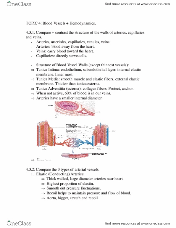
Describe the general structure and function of arteries and veins. General structure of arteries and veins: tunica interna. Simple squamous endothelium overlying a basement membrane and layer of fibrous tissue: tunica media. Usually thickest; smooth muscle, collagen, some elastic. Smooth muscle for vasomotion: tunica externa (tunica adventitia) Lower blood pressure of 10 mmhg (little fluctuation) Thinner walls, less muscular and elastic tissue. Valves aid skeletal muscles in upward blood flow. Veins with thin walls, large lumens and no smooth muscle. Describe the structure and function of the different types of arteries. Elastic arteries - are the largest arteries with examples including pulmonary a, aorta and common carotid a. Tunica media consists of perforated sheets of elastic tissue, alternating with thin layers of smooth muscle, collagen and elastic fibres. Expand during systole, recoil during diastole - lessens fluctuations in blood. Recoil in diastole keeps blood flowing while ventricles relax and prevents pressure blood pressure dropping too low.