DOH100 Lecture Notes - Lecture 1: Papillary Muscle, Interatrial Septum, Pulmonary Valve

Document Summary
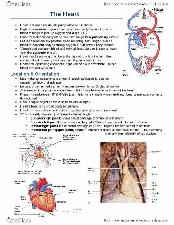
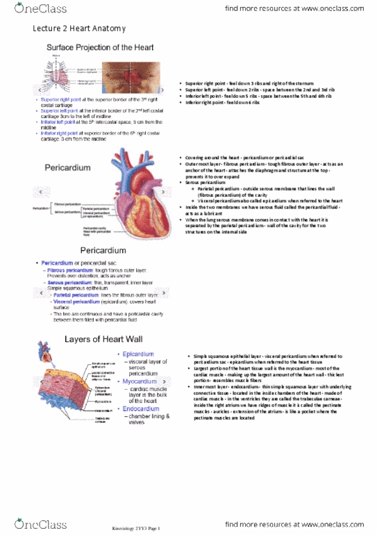
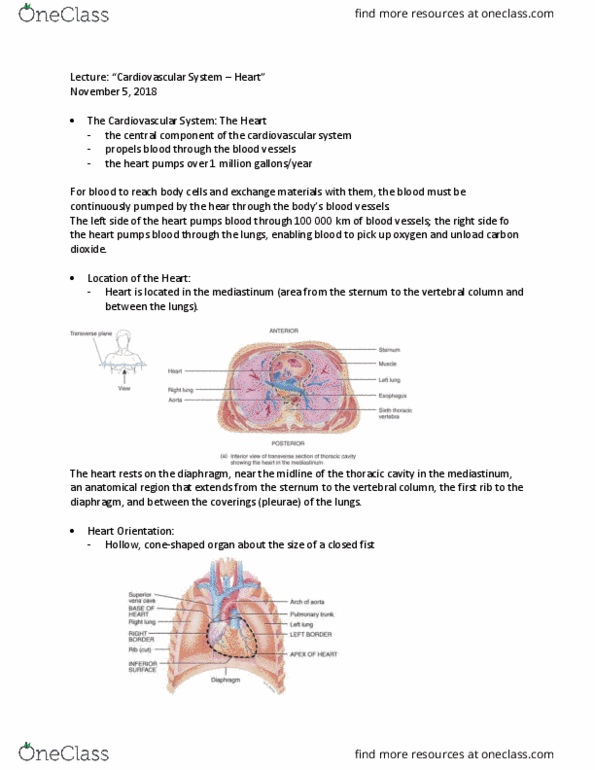
Describe the general location, size, and shape of the heart. Location: mediastinum, between lungs, resting superiorly on the diaphragm. Base - broad superior portion of heart. Apex - inferior end, tilts to the left, tapers to point (most of heart on left) 13 cm from base to apex and. A double walled sac that encloses the heart. Name & describe the 3 layers of the heart wall: epicardium. Fibrous skeleton - network of collagenous and elastic fibers which provides structural support and attachment for cardiac muscle. Electrical nonconductor, important in coordinating contractile activity: endocardium. Name, describe, identify & state the function, of the 4 heart chambers. Receive blood returning to heart from superior and inferior vena cava (right atrium) & pulmonary veins (left atrium) Ear-like extensions on the anterior surface of each atria are called auricles. Pump blood into pulmonary trunk (right ventricle) or ascending aorta (left ventricle)