BMSC11002 Lecture Notes - Lecture 8: Limbic System, Commissural Fiber, Body Fluid


1 | P a g e
WEEK 8: CENTRAL NERVOUS SYSTEM
13.1 THE CENTRAL NERVOUS SYSTEM: BRAIN, SPINAL CORD, AND PROTECTIVE STRUCTURES
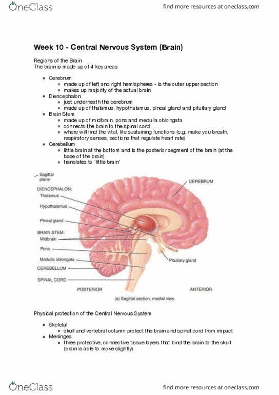
• the brain consists of 4 major parts:
o the cerebrum – is the largest part of the brain
o the diencephalon – contains the thalamus and hypothalamus
▪ provides a structural connection between the cerebrum and the brain stem
o the brain stem – consists of the midbrain, pons, and medulla oblongata
o cerebellum – posterior to the brain stem
• the spinal cord extends inferiorly from the medulla oblongata to the level of the first or second
lumbar vertebra
SKELETAL PROTECTION
• the first layer of protection for the central
nervous system is the hard bony skill and vertebral
column
• the brain is encased within the cranial cavity of
the skull, formed by the cranial bones
• the spinal cord is located within the vertebral
canal of the vertebral column
• the vertebral foramina of all the vertebrae,
stacked one on top of the other, form the vertebral
canal
• the surrounding skull and vertevral column
provide a strong, protective shelter against damage
find more resources at oneclass.com
find more resources at oneclass.com

2 | P a g e
MENINGES
• the meninges are three protective, connective coverings that lie betweent he bony encasement
of the skull and vertebral column and the brain and spinal cord
o the cranial meninges surround the brain and are conitinuous with the
o spinal meninges, which surround the spinal cord
• they have the same basic structure
o dura mater, which is composed of dense, irregular connective tissue
▪ the dura mater of the brain adheres directly to the periosteum of the interior
surface of the cranial bones
▪ epidural space exists between the dura mater and the wall of the vertebral canal
▪ the spinal cord is protected not only by the strong dura mater, but also by a cushion
of fat and connective tissue located within the epidural space
▪ three extensions of the dura mater separate portions of the brain
• the falx cerebri – separates two hemispheres of the cerebrum
• the falx cerebelii – separates two hemisphers of the cerebellum
• the tentorium cerebelli – separates the cerebrum from the cerebellum
find more resources at oneclass.com
find more resources at oneclass.com

3 | P a g e
▪ the dura mater of the brain contains dural sinuses, spaces within the dura mater
such as the superior sagittal sinus, which drains blood from the brain and delivers
it to the internal jugular veins of the neck
o the arachnoid mater, an avascular covering, named due to its spider-web-like
appearance of delicate collagen fibres and some elast fibres
▪ between the dura mater and arachnoid mater is the subdural space, which contains
interstitial fluid
o the innermost meningeal membrane is the pia mater, a thin, transparent connective
tissue layer that adheres to the surface of the brain and spinal cord
▪ contains interlacing bundles of collagen fibres and some fine elastic fibres
▪ triangular-shaped membranous extensions of the pia mater called dentriculate
ligaments project laterally from the spinal cord and fuse with the arachnoid mater
and inner surface of the dura mater
13.2 CNS BLOOD FLOW AND CEREBROSPINAL FLUID (CSF)
find more resources at oneclass.com
find more resources at oneclass.com
Document Summary
13. 2 cns blood flow and cerebrospinal fluid (csf) The ventricular system is a set of four interconnected cavities (ventricles) in the brain and the location of csf production. This system is continuous with the central canal of the spinal cord. The system comprises four ventricles: right and left lateral ventricles (the first and second ventricles) third ventricle fourth ventricle. The cavities of the cerebral hemispheres are called lateral ventricles or first and second ventricles. These two ventricles open into the third ventricle by a common opening called the foramen of monro. Csf is produced by modified ependymal cells of the choroid plexus found in all components of the ventricular system except for the cerebral aqueduct and the posterior and anterior horns of the lateral ventricles. The brain and spinal cord are covered by a series of tough membranes called meninges, which protect these organs from rubbing against the bones of the skull and spine.