PHTY100 Lecture 2: Arthrology and the hip joint


Introductory Arthrology and the Hip Joint
2.1 Define the term joint
A joint is a union between two or more parts of the skeleton.
2.2 Classify joints using the following criteria- relative amount of movement available and
structure:
•Fibrous (suture, syndesmosis)
Fibrous material joins the bone ends together. The amount of movement available depends on the
length of the fibres.
Suture:
-only found in the skull
-bones are linked by short fibres of connective tissue
-little to no movement
Syndesmosis:
-bones are linked by longer bands of connective tissue
-slightly moveable
•Cartilaginous (primary or synchondrosis and secondary or symphysis)
Cartilaginous material joins the bone ends together. The amount of movement available depends
on the
type of cartilage.
Synchondrosis:
-found where 2 bone growth centres in a developing bone remain separated by a layer of
cartilage
-these joints allow for bone growth and eventually completely ossify
-immoveable
Symphysis:
-where a pad/disc of fibrocartilage binds 2 bones together
-found in the mid-line of the body
-slightly moveable
•Synovial
Bone ends are shaped to fit one another, and covered with articular cartilage, allows the bones to
slide on one another.
Joint cavity contains synovial fluid (a lubricant) which assists in frictionless movement of bones.
Fibrous joint capsule unites the bones and maintains the joint cavity.
Synovial membrane lines the fibrous capsule and produces synovial fluid.
2.3 Further classify synovial joints using the following criteria:
•Degree of freedom (uniaxial, biaxial, multiaxial)
-uniaxial – one pair of movements
-biaxial – two pairs of movements
-multiaxial – three pairs of movements
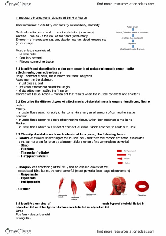
•Structure (hinge, pivot, ellipsoid, condyloid, saddle, ball and socket, plane)
•Uniaxial
-Hinge
-Pivot
•Biaxal
-Ellipsoid
-Condyloid
-Saddle
•Multiaxial
-Ball and socket
-Plane
2.4 List and identify examples of each type of joint mentioned in objectives 2.2 and 2.3
(across both upper limb and lower limb)
•Fibrous

-Suture- only found in skull
-Syndesmosis- wrist between ulna and radius and ankle between tib fib
•Cartliganious
-Synchondrosis- epiphyseal growth plates
-Symphysis- joints between bodies of adjacent vertebrae, joint between bodies of pubic bones
•Synovial
-Hinge (uniaxial)- elbow
-Pivot (uniaxial)- 1st-2nd cervical vertebrae
-Ellipsoid (biaxial)- radiocarpal joint
-Condyloid (biaxial)- metacarpophalangeal joints
-Saddle (biaxial)- carpometacarpal joint of thumb
-Ball and socket (multiaxial)- hip joint
-Plane (mutliaxial)- acromioclavicular joint, tarsal bones in foot
2.5 In individual synovial joints, assign the terms male and female to describe the shape of
the articular surfaces
The ‘male’ is the bone that fits into the ‘female bone’.
2.6 List the functions of ligaments
Ligaments are bands of fibrous tissue that occur at all types of joints.
Function:
-act as mechanical constraints (mechanical function)
-prevent unwanted movement
-limited allowed movement
-sensory organs - proprioception
2.7 Understand the classification of ligaments as capsular, extracapsular and intracapsular
•capsular – reinforce the capsule
•extracapsular – lie outside the capsule
•intracapsular – lie inside the capsule
2.8 Identify examples of capsular, extracapsular and intracapsular ligaments
•Capsular- illiofemoral ligament, pubofemoral ligament, ischiofemoral ligament
•Extracapsular- medial collateral ligament (MCL), lateral collateral ligament (LCL),
•Intracapsular- anterior cruciate ligament (ACL), posterior cruciate ligament (PCL), ligaments
teres
2.9 Understand the structure of and list the functions of articular disks
Articular disks are pads of fibrocartilage that are situated between the articular surfaces of some
synovial joints.
Function to:
-act as shock absorbers
-aid mechanical fit between articular surfaces
-restrain movement
-assist lubrication
-permit different movements to occur simultaneously in the one joint
2.10 Understand the structure of and list the functions of bursae
The bursae is fluid filled sacs around many synovial joints. Potential rather than
actual spaces.
The function is to reduce friction as structures slide on one another. Therefore located between
layers of muscles and where muscles and tendons overlie bony prominences.
2.11 Describe the 3 principal axes of movement about synovial joints
•anteroposterior axis – front to back (abduction/adduction, lateral flexion)
•transverse axis – side to side (flexion/extension)
Document Summary
A joint is a union between two or more parts of the skeleton. 2. 2 classify joints using the following criteria- relative amount of movement available and structure: fibrous (suture, syndesmosis) The amount of movement available depends on the length of the bres. Bones are linked by short bres of connective tissue. Bones are linked by longer bands of connective tissue. Slightly moveable: cartilaginous (primary or synchondrosis and secondary or symphysis) The amount of movement available depends on the type of cartilage. Found where 2 bone growth centres in a developing bone remain separated by a layer of. These joints allow for bone growth and eventually completely ossify. Where a pad/disc of brocartilage binds 2 bones together. Found in the mid-line of the body. Bone ends are shaped to t one another, and covered with articular cartilage, allows the bones to slide on one another. Joint cavity contains synovial uid (a lubricant) which assists in frictionless movement of bones.