BIOL126 Lecture Notes - Lecture 9: Heart Valve, Heart Murmur, Depolarization

10 Jun 2018
School
Department
Course
Professor

The Heart
Cardiac physiology:
-As atria relax, ventricles contract
-2 types cardiac muscle cells:
-conduction system cells, contractile cells
-Conduction- smaller diameter, control, coordinate heart beat
-Contractile- powerful force propels blood
-Sinoatrial node (SA)- pacemaker of heart (AP generated here)
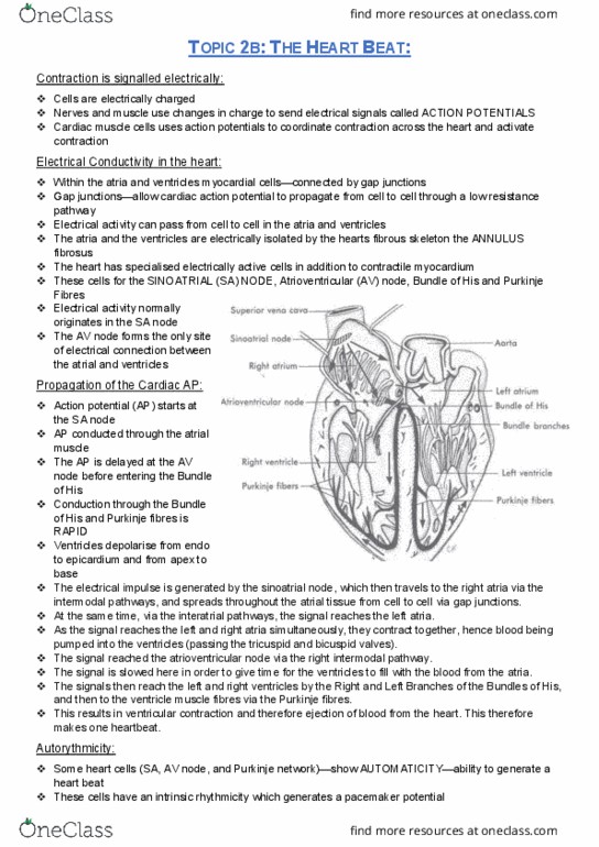
Conducting system:
-(coordinates contracting)
-Nodal system
-Contraction lags behind AP- delay time for Ca+ ions to enter sarcoplasm
-Cardiac muscle needs high Ca2+
-Two nodes- Sa (top of RA), atrioventricular node
Purkinje fibres:
-triggers contraction of the ventricles from the apex up towards the arteries
-SA is pacemaker as has faster depolarisation rate - gets hear beat/ heart rate
-doesn’t have set resting membrane potential
-AV can take over if SA damaged however rate slowers
AP in cardiac muscle cells:
1. rapid depolarisation
2. Plateau (fairly long)
3. Repolarisation
-once AP generated = really quick depolarisation= voltage gated Na+ chemicals open, increased
charge
Conduction pathway:
-delay occurs as diameter of conduction cells bringing signal to AV node is larger than the AV
nodal itself- if it didn't have this delay the atria wouldn’t of finished contracting and be able to
recover when the ventricles start contracting- delay in contract
Electrocardiogram (ECG):
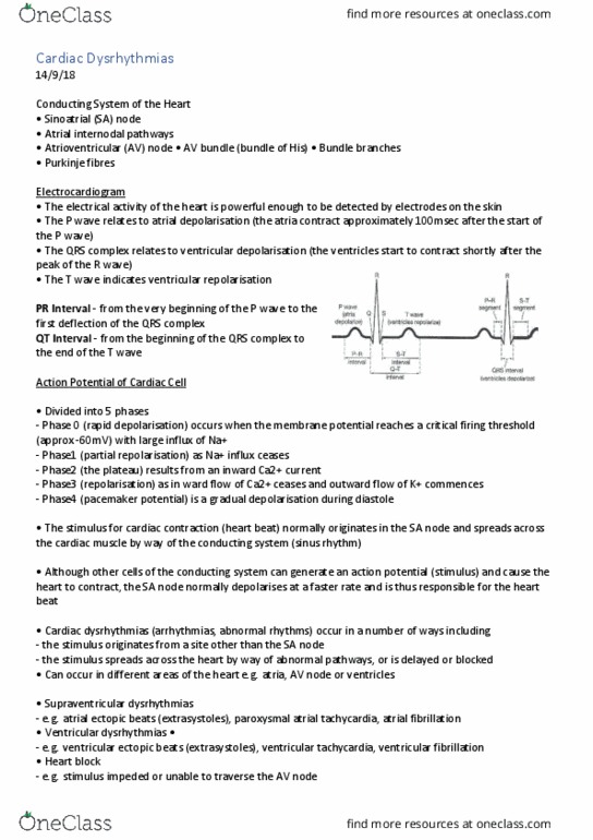
-Normal sirus rhythm comprises P wave, QRS complete, T wave
-if problem with P wace- likely problem with atrium
-atrial depolarisation= p face- atrial contraction
-atrial repolarisation = QRS complete- atrial relaxation
-ventricle depolarisation = QRS complete with R wave= ventricle contraction
-Ventricle repolarisation = T wave = ventricle relaxation
AP in cardiac muscle cells:
-Contraction process much larger for cardiac than skeletal muscle cells
-Rapid depolarisation = voltage goes Na+ channels open
-Plateau = Na+ channels start to close, Ca2+ open
-Repolarisation = Ca2+ start to close, K+ open
-Ca2+ triggers release of extra Ca2+ ions from sarcoplasmic relaxation
Heart sounds:
-Heart murmur = problem with AV valve function
-Lubb = closure of AV valves, lasts a bit longer- sound from blood lifting valves
-Dupp = losure of SL valves, blood lifting valves
-Abnormal sounds = blood flowing into ventricles + atrial contraction
find more resources at oneclass.com
find more resources at oneclass.com
Document Summary
Conduction- smaller diameter, control, coordinate heart beat. Sinoatrial node (sa)- pacemaker of heart (ap generated here) Contraction lags behind ap- delay time for ca+ ions to enter sarcoplasm. Two nodes- sa (top of ra), atrioventricular node. Triggers contraction of the ventricles from the apex up towards the arteries. Sa is pacemaker as has faster depolarisation rate - gets hear beat/ heart rate. Av can take over if sa damaged however rate slowers. 1. rapid depolarisation: plateau (fairly long, repolarisation. Once ap generated = really quick depolarisation= voltage gated na+ chemicals open, increased charge. Normal sirus rhythm comprises p wave, qrs complete, t wave. If problem with p wace- likely problem with atrium. Atrial repolarisation = qrs complete- atrial relaxation. Ventricle depolarisation = qrs complete with r wave= ventricle contraction. Ventricle repolarisation = t wave = ventricle relaxation. Contraction process much larger for cardiac than skeletal muscle cells. Rapid depolarisation = voltage goes na+ channels open.