ALHT106 Lecture Notes - Lecture 7: Septum Secundum, Endocardial Tubes, Interventricular Septum


Development of the cardiovascular system:
-The embryonic vascular system commences development in the third week
-vessels within the developing foetal membranes (placenta, yolk sac)
-vessels within the developing embryo
-formation of the embryonic heart
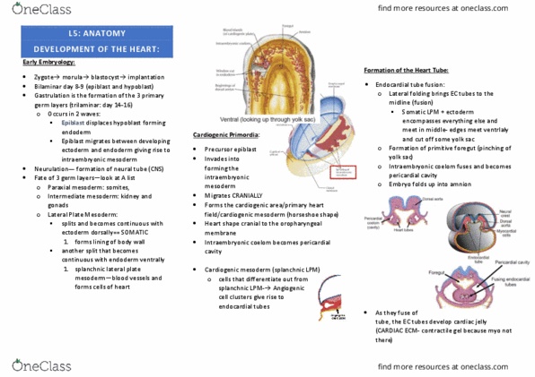
Development of the heart:
-The left and right endocardial tubes form within the lateral splanchnic
mesoderm, cephalic to the buccopharyngeal membrane
-Lateral folding causes the two tubes to meet in the midline and fuse to form a
single endocardial heart tube
-Cephalocaudal folding causes the devleloping heart to move caudally, and
come to lie caudal to the buccopharygneal membrane
-The endocardial tube becomes surrounded by a thickened layer of mesoderm
which will form; myocardium and visceral pericardium
-At each end of the endocardial tube are blood vessels that connect the
developing heart to the reminder of the embryo and the foetal membrane
(more soon)
-The heart begins to beat in the third week
-Rapid growth of the endocardial tube causes; appearance of bulges along the
tube length, bending of the tube
Partitioning of the heart:
-Partiotions within the heart form between days 27 and 37
-At the junction of the developing atria and ventricles the mesoderm proliferates on the dorsal
and ventral walls
-Eventually these proliferations will meet and fuse (forming the septum intermedium)
-Simultaneously a septum grows from the roof of the developing atrium- septum premium
-Grows downward to fuse with the septum intermedium
-Upper part of the septum premium will degenerate forming the foramen secundum
-Second septum also grows from the right side of the roof of the atrium-s septum secundum
-A muscular projection grows upward from the floor of the ventricle- ventricular septum
-The ventricular septum will fuse with the septum intermedium
-Separation of the left and right ventricles
-Each ventricle communicates with its respective atria
-The left and right atria are in communication
-Distal bulbous cordis and trunks arterioles become partitioned into an aorta and a pulmonary
trunk (artery) by the spinal aorticopulmonary septu
-Seperation of the left and right ventricles
-Each ventricle communicates with its respective atria
-The left and right atria are in communication
-The right ventricle communicates with the pulmonary trunk, the left ventricle with the aorta
Foetal circulation: Adult circulation:
Atypical development of the heart:
find more resources at oneclass.com
find more resources at oneclass.com

-Persistent spatial defects- atrial, ventricular
-Persisten trunque arteriosus
-no division into aorta and pulmonary trunk
-Single artery arising from the heart
-Patent ductus arteriosus
-aortic blood passes into the pulmonary circulation
-Increased pressure in the pulmonary circulation
Development of the respiratory system:
-Development commences in the fourth week
-Outgrowth from the foregut- laryngotracheal tube
-Initially in contact with the foregut but will eventually
seperate from the foregut
-During the fifth week the caudal part of the laryngotracheal tube divides into the left and right
lung buds
-right lung bud more in line with the developing trachea, maintained throughout adult life
-Left lung bud divides into two branches, the right lung bud divides into three branches
-Lung buds and developing bronchi are endoderm with a covering of splanchnic mesoderm
-endoderm becomes respiratory epithelium
-mesoderm becomes cartilage and muscle of ironical tree, plus visceral pleura
-Push into the space that will become the pleural cavity
-Progressive branching of the bronchi continues, by the sixth prenatal month there will be 17
generation of bronchi
-A further six generations will be formed to reach full development
-Alveolar development occurs from the fifth month and continues after birth
-Cuboidal cells of the terminal branches flatten to form alveoli
-Surfactant produced by selected alveolar cells from the 30th week
Development of the diaphragm:
-Forms from a mesoderm structure- septum transversum
-Fusion of the mesoderm from the hypomeres of c3, C4 and C5 (not phrenic nerve root value)
-Pushed caudally by descent of heart from the neck into the thorax associated withe
cephalocaudal folding
Development of the head and neck:
Pharyngeal arches:
-Five pairs of pharyngeal arches develop on either side of the foregut,
each is:
-covered withe externally by ectoderm
-lined with endoderm
-filled with mesoderm
-each becomes associated with a cranial nerve
-Between adjacent arches externally- pharyngeal clefts, internally-
pharyngeal pouches
-Ectoderm covering
-skin of face and neck
-Endoderm lining
-lining of tympanic cavity, tonsils, some glands
-Mesoderm core
-bone and skeletal muscle
-Each arch grows forward and medically and will eventually fuse on the anterior midline
Pharyngeal arches- innervation:
-Cranial nerves grow into arches:
-- I - CN V
-II - CN VII
find more resources at oneclass.com
find more resources at oneclass.com

-III - CN IX
-IV - CN X
-V - not in humans
-VI - CN X
Development of the face:
-The first pharyngeal arch will develop into the structures of the face (note the innervation)
-division into a maxillary process, a mandibular process and a frontonasal process
-migrate forward and meet in the midline forming the mandible, maxilla, lips, nose, palate and
cheeks accomodating the mouth and nostrils
-forms the muscles of mastication
Development of other head and neck structures:
-The second pharyngeal arch will become:
-muscles of facial expression
-part of the external ear
-The third pharyngeal arch will become:
-hyoid bone
-some muscles of the pharynx
-The fourth pharyngeal arch will become
-hyoid bone, larynx
-muscles of the soft palate, pharynx and larynx
Microbiology:
Topics:
Types of microorganisms
Factors that affect bacterial growth and survival
Gram staining
Normal flora
Transmission of microorganisms
Requirements for disease
Pathogenic properties
Methods of control
What is microbiology:
The study of microscopic organisms (microorganisms)
Microorganisms: living organisms that need a microscope to study them
Cells are generally less complex
microorganisms are everywhere and mostly unseen
Introduction:
-microorganism- first life on earth- 3.8 billion years ago
-microorganisms created the biosphere that allowed multicellular organisms to evolve
-Multicellular organisms evolved from microorganisms
-Most of the physiological, metabolic and genetic diversity belongs to the microbial world
-EVERYWHERE!- 50% of biomass is made of microorganisms
find more resources at oneclass.com
find more resources at oneclass.com
Document Summary
The embryonic vascular system commences development in the third week. Vessels within the developing foetal membranes (placenta, yolk sac) The left and right endocardial tubes form within the lateral splanchnic mesoderm, cephalic to the buccopharyngeal membrane. Lateral folding causes the two tubes to meet in the midline and fuse to form a single endocardial heart tube. Cephalocaudal folding causes the devleloping heart to move caudally, and come to lie caudal to the buccopharygneal membrane. The endocardial tube becomes surrounded by a thickened layer of mesoderm which will form; myocardium and visceral pericardium. At each end of the endocardial tube are blood vessels that connect the developing heart to the reminder of the embryo and the foetal membrane (more soon) The heart begins to beat in the third week. Rapid growth of the endocardial tube causes; appearance of bulges along the tube length, bending of the tube. Partiotions within the heart form between days 27 and 37.