ALHT106 Lecture Notes - Lecture 1: Dermis, Hair Follicle, Cholecalciferol


1. Discuss the structure and function of the integument system
2. Summarise the changes that occur to the integument system across the lifespan
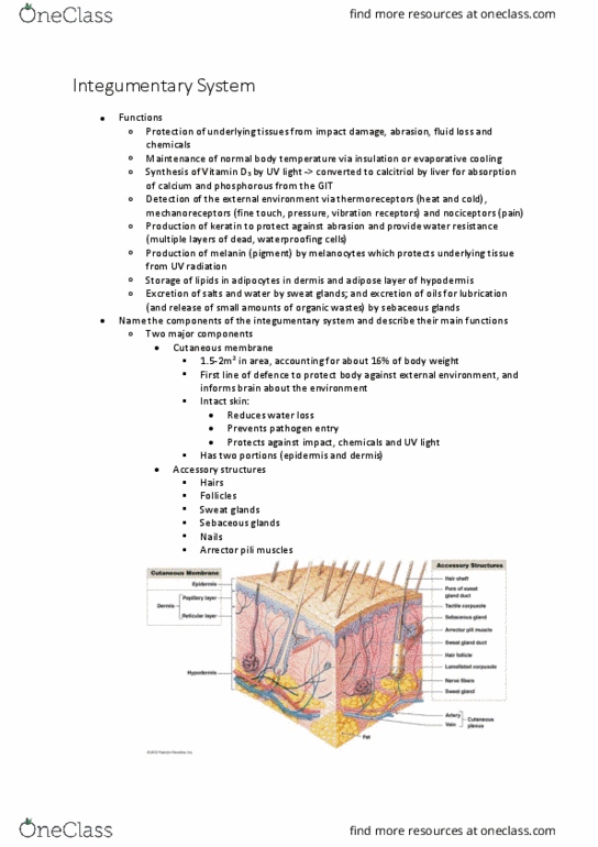
•Name the components of the integumentary system and describe their main functions
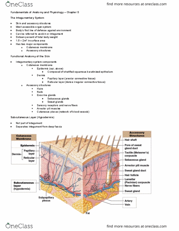
Integumentary system:
-two major components:
-cutaneous membrane
-Accessory structures
-1.5-2m2 in area, accounting for about 16% of body weight
-first line of defence to protect body against external encironment, and informs brain about the
environment
-intact skin reduces water loss, prevents pathogen entry, and protects against impact, chemicals
and UV light
-Accessory structures: hair, follicles, sweat glands, sebaceous glands, nails; arrestor pili muscles
-cutaneous membrane has two portions: epidermis and dermis
-Epidermis comprised from stratified squamous epithelium; dermis from areolar/loose connective
tissue; and dense, irregular connective tissue
-hypodermis (subcutaneous adipose layer) separates integument from deep fascia and other
organs
-connective tissue fibres of hypodermic interweave with those of the dermis- hold tissue layers
together; epidermal ridges and dermal papilla increase SA for interconnections between
epidermal and dermal layers
Integumentary system functions:
1. protection of underlying tissues from impact damage, abrasion, fluid loss and chemicals
2. Maintenance of normal body temp. via insulation or evaporative cooling
3. Synthesis of vitamin D3 by UV light- converted to calcitriol by liver for absorption of calcium and
phosphorus from the GIT
4. detection of the external environment via thermoreceptors (heat and cold), mechanoreceptors
(fine touch, pressure, vibration receptors) and nociceptors (pain)
5. production of keratin to protect against abrasion and provide water resistance (multiple layers
of dead, waterproofing cells)
6. production of melanin (pigment) by melanocytes which protects underlying tissue from UV
radiation
7. storage of lipids in adipocytes in dermis and adipose layer of hypodermic
8. Excretion of salts and water by sweat glands; and excretion of oils from lubrication (and release
of small amounts of organic wastes) by sebaceous glands
•Identify the 3 layers of skin (and their component parts), and relate the structure of each
layer to its function
Accessory structures:
Two types of sweat glands:
1. Apocrine- pubic and axillary regions and around nipples; activated at puberty; secretes sweat
into hair follicles - odour since bacteria use it for nutrition
2. Merocrine- secrete onto skin surface; more numerous and widely distributed’ palms and soles
have highest numbers; comprised of water, electrolytes (mainly Nail), organic compounds, and
an antibiotic peptide (dermacidin)
-cool skin surface to reduce body temp.; excrete water, electrolytes and some drug metabolises;
dilutes harmful chemicals on skin; discourages microbial growth by flushing off skin or via
dermacidin
-both contain anti-bacterial molecules and help cool skin
•Summarise the structure and function of the epidermis, dermis and hypodermic
Epidermis:
-stratified squamous epithelium provides mechanical protection and prevents entry of
microorganisms
find more resources at oneclass.com
find more resources at oneclass.com

-layers or strata present: thin skin has four layers, thick skin on soles, palms and fingertips has
fifth layer (Stratum lucid)
-all epithelium is avascular, so living tells are in the basal layer by the basement membrane or
dendritic cells in second layer
-mitosis provides continuous replacement when the dead cells at the surface are worn away
-cells die, progressively flatten and fill with keratin as they move towards the surface- 15-30
layers of dead waterproofing cells at top
Epidermal cell types:
1. Basal cells: the germinative cells which undergo mitosis to replace the continual loss of upper
layer cells
2. Keratinocytes: most common epithelial cell; filled with keratin protein; flatten and die as they
progress outwards. outermost layer is 15-30 layers of dead keratinocytes tightly connected by
desmosomes
3. Melanocytes: pigment-producing cells in basal layer (skin and hair colour)
4. Dendritic (langerhans) cells: in 2nd layer; defend against microbes that penetrate skin; and
superficial skin cancers
5. Merkel cells: tactile cells scattered in basal layer (of skin without hair)
Roles of skin pigmentation:
-two pigments produced by melanocytes and transferred to keratinocytes; around 1000
melanocytes/mm2 in most skin
-Melanin: brown, yellow-brown to black; protects against UV damage
-Different forms of melanin- dark brown, yellow brown and red hair; hormones and environmental
factors also influence hair colour
-Carotene: orange-yellow; also stored in fat/adipose cells; can be converted to vitamin A
-Melanocytes increase pigment production in response to sun exposure, but too slowly to prevent
sunburn (peak 10 days post sun exposure)
Role of dermal circulation:
-Oxygenated blood looks bright red since Hb bound to to oxygen- oxyhemoglobin. As oxygen is
released and more Hb formed, blood gets a darker red-blue colour- red in arterial blood and blue
in venous blood as seen through the skin
-vasodilation of blood vessels in dermis increases blood flow through capillaries- pink coloured
skin and greater conduction of heat through the skin i.e greater heat loss
-Vasoconstriction of skin blood vessels has the opposite effect- heat conservation and pale skin
-skin easily observed and can aid diagnosis of body conditions
Disease and skin colour:
-a drop in blood supply to a tissue turn it pale/white (ischaemia); longer-term drop in blood flow-
cyanosis (blue colour) and hypoxia- cell death
-Jaundice: liver and/or kidneys cannot excrete enough bile or bile pigments- build-up in body
fluids, then sclera and skin
-tumours affecting the anterior pituitary can increase MSH- secretion of large amounts of
melanin- bronzing or darkening of skin
-Addison’s disease: anterior pituitary sects large amounts of ACTH (structurally similar to MSH),
so melanocytes respond by increasing secretion of melanin
-Vitiligo: suspected to be autoimmune where antibodies attack melanocytes; occurs in 1% of
population; loss of melanocytes and melanin
Dermis:
-Two layers:
-superficial papillary layer of areolar tissue; capillaries, lymphatic and sensory neurons
-deeper reticular layer of dense; irregular connective tissue; collagen, elastin; blood vessels;
nerve fibres; contain sweat and sebaceous glands; contain sensory receptors
-Collagen: great tensile strength; some give but resists stretching, pulling and twisting
find more resources at oneclass.com
find more resources at oneclass.com

-Elastin: gives stretch and recoil ability to skin
-Skin turgor: water present which gives resilience and flexibility to skin
-Alignment of the collagen fibres along tension lines allows skin to resist forces applied during
normal movement
•Explain the functions of accessory structures to the skin (hair and hair follicles, erector
muscles, sebaceous glands, sweat glands, nails) and why skin is pigmented
-Hairs: non-living (keratin + cuticle) grow from hair follicles in epidermis- grow down into dermis-
extend above skin surface
-protect from UV damage; help cushion light impact; reduce insect and pathogen entry e.g. on
head, in nostrils, eyelashes, eyebrows
-Important as sensory receptors (sensory neurons around base of every hair follicle- feel
movement in a single hair shaft- great sensitivity)
-around 2.5 million hairs on most of body- 25% or 500,000 on head
-Contraction of arrestor pili muscles (smooth muscle) - hairs upright
-Follicle: surrounded by connective tissue then sensory neutron (root hair plexus)
-Sebaceous glands: oil glands; discharge oily, lipid secretion (sebum) into hair follicles, and onto
skin
-Sebrum: comprised from triglycerides, cholesterol, proteins, electrolytes; inhibits bacterial
growth; lubricates and protects keratin of the hair shaft
-Sebaceous follicles discharge serum directly onto epidermal surface; located on face, back,
chest, nipples and external genitalia (only difference is where they are being secreted)
Nails:
-Keratin epidermal cells
-protect exposed tips of digits
-Help digit withstand distortion when subjected to mechanical stress- allows back force for tissue/
stress or pressure be able to do things e.g. pick things uo
Skin as a sense organ:
-sensory receptors: specialised cells which activate sensory neurons when stimulated
-simplest receptors: dendrites of sensory neurons e.g. nociceptors
-nociceptors detect pain; mechanoreceptors detect physical distortion; thermoreceptors detect
external temp. changes
-skin has a rich supply of sensory receptors: anything in contact with the skin activates sensory
pathways
Skin sense receptors:
-Nociceptors: respond to extremes of temperature; dissolved chemicals, including those released
by damaged cells; mechanical/physical damage; infection; inflammation; ischaemia
-Stimualtion of dendrites of nociceptor activates sensory neuron. Nociceptors do not show
adaption
-Thermoreceptors: free nerve endings in dermis (like nociceptors)
-no structural differences between hot and cold receptors. 3-4x more cold than hot receptors.
very active while temperature changing, but adapt quickly
-Mechanoreceptors: sensitive to stimuli which distort cell membranes
•Explain the functions of the main types of receptors in skin: nociceptors,
mechanoreceptors and thermoreceptors
Tactile receptors/ Mechanoreceptors
-detect touch, pressure and vibrations (closely related sensations)
-touch provides information about shape or texture
-pressure provides information about mechanical/physical distortion
-vibration provide information about pulsing pressure
-receptors may be specialised e.g. rapidly adapting tactile receptors best suited for vibration
find more resources at oneclass.com
find more resources at oneclass.com
Document Summary
Discuss the structure and function of the integument system, summarise the changes that occur to the integument system across the lifespan, name the components of the integumentary system and describe their main functions. 1. 5-2m2 in area, accounting for about 16% of body weight. Rst line of defence to protect body against external encironment, and informs brain about the environment. Intact skin reduces water loss, prevents pathogen entry, and protects against impact, chemicals and uv light. Accessory structures: hair, follicles, sweat glands, sebaceous glands, nails; arrestor pili muscles. Cutaneous membrane has two portions: epidermis and dermis. Epidermis comprised from strati ed squamous epithelium; dermis from areolar/loose connective tissue; and dense, irregular connective tissue. Hypodermis (subcutaneous adipose layer) separates integument from deep fascia and other organs. Connective tissue bres of hypodermic interweave with those of the dermis- hold tissue layers together; epidermal ridges and dermal papilla increase sa for interconnections between epidermal and dermal layers. Cool skin surface to reduce body temp.