PHIL 2130 Chapter Notes - Chapter 2.2: Macula Of Retina, Fovea Centralis, Sensory Nerve

6 May 2020
School
Department
Course
Professor
Document Summary
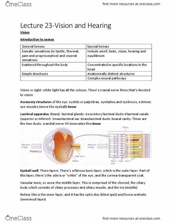
Retina also contains rods, cones and photopigment molecules. Fovea centralis is a small depression in the center of the macula lutea (center of the eyeball) and it contains only cones-know. Light touches the pigmented layer first though (nerve impulses and light travel in opposite directions)-know. Cones: three types-red, green and blue (these cones sense colour) Outer segment contains photopigments, and the transduction of light energy into receptor potential occurs here. The inner segment contains the nucleus, golgi complex and mitochondria. Ganglion cells in retina of each eye join to form optic nerve. Supply extrinsic eye muscles to control movements of eyeball and upper eyelid. Smallest of the 12 cranial nerves, originiates in the midbrain. Sensory portion extends from the taste buds of the anterior two thirds of the tongue. Motor portion arises from the pons and deals with facial expression. Light adaptation (dark to light) is fast, while dark adaptation (light to dark) is slow.