PSY290H5 Chapter Notes - Chapter 5: Transcranial Magnetic Stimulation, Positron Emission Tomography, Diffusion Mri

Document Summary
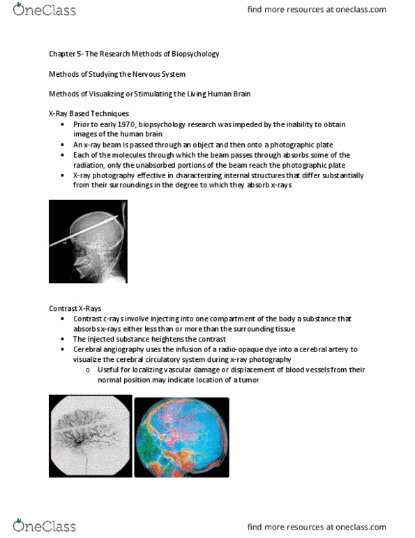
Digital atlas of gene expression patterns in the mouse/human brain. Injecting a substance into a compartment of the body so that it absorbs more or less than the rest of the surrounding tissue. Heightens the contrast between the structures and makes it easier to see: e. g. cerebral angiography: put a radio-opaque dye into a cerebral artery to visualize the cerebral circulatory system during x-ray. Functional mri (fmri) fmri advantages over pet: both structural and functional info in same image, nothing has to be injected into the volunteer, spatial resolution is better (but poorer temporal resolution, 3d image of entire brain. Since tracts (bundles of axons) are the major routes of rapid water diffusion in the brain diffusion tensor imaging provides an image of major tracts. Temporarily shuts off a cortical area by creating a magnetic field under a coil positioned next to the skull and see how cognition and behaviour are effected.