BPK 325 Chapter Notes - Chapter 11: Detrusor Urinae Muscle, Membranous Urethra, Prostatic Urethra

16 Aug 2020
School
Department
Course
Professor
Document Summary
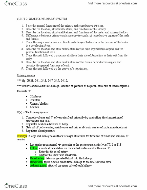
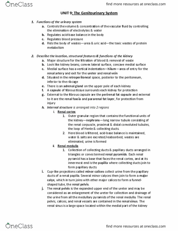
Kidneys sit on the posterior abdominal wall, have concave border facing medially. Sit around the t12-l3 vertebrae level, right kidney sits slightly lower (right lobe of liver pushes it down a bit) Surrounded by tough renal capsule (dense, irregular ct) Superficial to fat pads renal fascia (anchors kidneys posterior abd walls, keeps it in place) Deep to capsule renal fat pads (absorbs shock) Renal pyramids (pyramid-shaped structures near center) area makes up renal medulla. Point of pyramid = renal papillae drains bundles of collecting ducts into calyces (cup-like structure) Calyces drain urine into renal pelvis then out through ureter. Renal cortex (seen on outer aspect of the kidney) Renal columns (fill spaces between the pyramids) Each lobe of kidney has pyramid and nearby associated regions of renal cortex. Renal hilum contains renal vessels, a nerve, the ureter (all on medial side) Functional unit of the kidney, ~1 mil units.