ANAT2111 Lecture Notes - Lecture 20: Transpyloric Plane, Renal Papilla, Renal Pelvis

21 Jun 2021
School
Department
Course
Professor
Document Summary
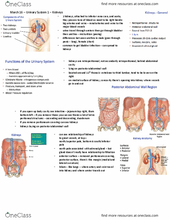
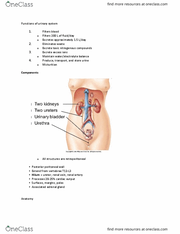
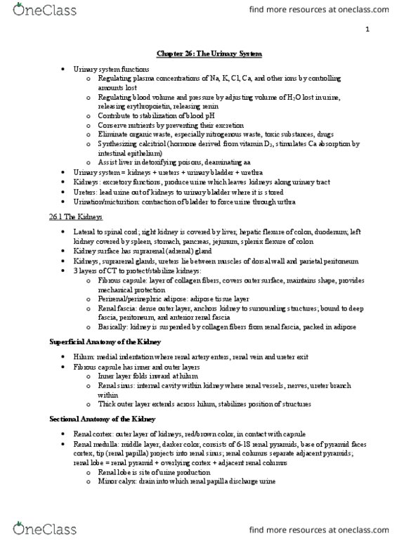
Maintaining electrolyte, acid/base, fluid balance and ionic composition. Secrete hormones (aid in it"s function) e. g. calcitriol and erythropoietin. Activation of vitamin d together with liver. Paired kidneys form urine ureter bladder (temporary storage sac for urine) urine expelled via the urethra. Kidneys (retroperitoneal) - length: t12 - l3 (10-12cm x 5-7cm) Passes through hilum of kidneys + pylorus of stomach. Right kidney sits slightly inferior to left due to liver. Hilum - contain renal artery + vein and ureter. Surrounded by three layers of connective tissue: Renal fascia: most external: anchor to wall. Renal pyramid: base cortex, apex (renal papilla) projects inwards. Minor calyx surrounds renal papilla urine is collected. Minor calyx drains major calyx form renal pelvis (funnel lead into ureter). 1 mill nephrons + thousands of ducts per kidney. Inside: blood is filtered; nutrients, water, essential electrolytes are reabsorbed, waste and excess substances are secreted. Urine passes collecting ducts minor. Descend retroperitoneally to abdomen enter pelvic cavity.