PSYC 273 Lecture Notes - Lecture 6: Oval Window, Middle Ear, Sound


Chapter 6 Notes
• Afferent= Ascending to brain. Bottom up processes.
• Efferent= Descending from brain. Top down processes.
• Phasic receptors: DO experience sensory adaptation. (stimulus phases out).
• Tonic receptors: do not experience sensory adaptation. (e.g. many pain signals).
Part I: Hearing and Balance
• Amplitude: a.k.a. intensity. The force that sound exerts per unit area, usually measured
as dynes per square centimeter. Corresponds to the volume of the sound. Measured in
decibels.
o Decibel: a measure of sound intensity, perceived as loudness.
o Perceived as loudness.
• Frequency: the number of cycles per second in a sound wave, measured in hertz.
o Hertz: cycles per second, as of an auditory stimulus.
o Perceived as pitch.
o Pitch: a dimension of auditory experience in which sounds vary from low to high.
• Pure Tone: a tone with a single frequency of vibration.
• Fundamental: the predominant frequency of an auditory tone or a visual scene.
o Harmonics: a multiple of a particular frequency called the fundamental.
• Timbre: the characteristic sound quality of a musical instrument, as determined by the
relative intensities of its various harmonics.
• Pitch and loudness are psychological properties, frequency and amplitude are physical
properties.
Each Part of the Ear Performs a Specific Function in Hearing
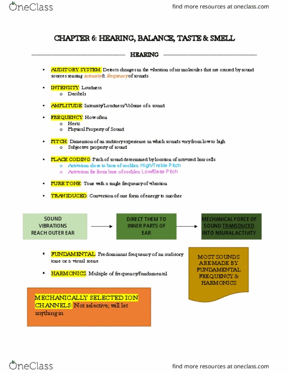
• Transduction: the conversion of one form of energy to another.
o Sound is transduced into neural activity.
• The external ear captures, focuses, and filters sound.
o Pinnae: the external part of the ear. Funnel sound waves into the second part of
the external ear. Strictly for mammals.
▪ The hills and valleys of the pinnae modify the character of sound that
reaches the middle ear.
• Ear Canal: the tube leading from the pinna to the tympanic membrane.
• The middle ear concentrates sound energies.
o Inner Ear: the cochlea and the vestibular apparatus.
o Middle Ear: the cavity between the tympanic membrane and the cochlea.
o Tympanic Membrane (Eardrum): the partition between the external ear and the
middle ear. Moves the Ossicles.
o Ossicles: 3 small bones that transmit vibration across the middle ear, from the
tympanic membrane to the oval window. The smallest bones in the body. Move
the oval window.
▪ Malleus: a middle ear bone that is connected to the tympanic
membrane.
▪ Incus: a middle ear bone situated between the malleus and the stapes.
find more resources at oneclass.com
find more resources at oneclass.com

▪ Stapes: a middle ear bone that is connected to the oval window. Like a
hammer on the oval window.
o Oval Window: the opening from the middle ear to the inner ear.
o Process:
▪ Sound waves strike the tympanic membrane and cause it to vibrate at the
frequency of that sound.
▪ Causes the Ossicles to move, concentrate and amplify the vibrations, and
focus the pressure towards the smaller oval window.
▪ Hits the oval window via the stapes.
o Protected with sound control to protect the ear through the contraction of 2
muscles when any loud sound happens. Help control intensity and self-made
noises. Reducing the vibration of the Ossicles.
▪ Tensor Tympani
▪ Stapedius
• The cochlea converts vibrational energy into neural activity.
o Cochlea: a snail shaped structure in the inner ear that contains the primary
receptor cells for hearing. Converts vibration to sound.
▪ Vestibular Canal= Scala Vestibuli: 1 of 3 principal canals running along
the length of the cochlea.
▪ Middle Canal= Scala Media: the central of the 3 canals inside the
cochlea, situated between the vestibular canal and the tympanic canal.
▪ Tympanic Canal= Scala Tympani: 1 of 3 principal canals running along the
length of the cochlea.
o Round Window: a membrane separating the tympanic canal from the middle
ear. If stapes pushes oval inward then round bulges out.
o Organ of Corti: a structure in the inner ear that lies on the basilar membrane of
the cochlea and contains the hair cells and terminations of the auditory nerve.
Turns sound into neural activity. Contains the basilar membrane.
▪ Axons are coming off of Organ of Corti along entire length.
• Each axon extends from specific location along membrane.
• In turn, each location corresponds to the specific frequency that
maximally displaces that location.
▪ Consists of 3 main structures.
• Hair Cells: one of the receptor cells for hearing in the cochlea,
named for the stereocilia that protrude from the top of the cell
and transduce vibrational energy in the cochlea into neural
activity.
• An elaborate framework of supporting cells.
• The terminations of the auditory nerve fibers.
▪ The base of the Organ of Corti
• Basilar Membrane: a membrane in the cochlea that contains the
principal structures involved in auditory transduction. Widens and
find more resources at oneclass.com
find more resources at oneclass.com

becomes more flexible toward apex, thus lower frequencies move
further along.
o Base: nearest the oval window. Narrow and stiff. (higher
frequency).
o Apex: at the end. Innermost of coil. Wider and more
flexible. (lower frequency).
• Process:
o Stapes move in and out due to sound waves hitting the ear
drum.
o Sets in motion waves in the fluid of the vestibular canal.
o Causes the basilar membrane to ripple (like shaking out a
rug). High frequencies have greater effect near the base
(where it is very narrow). Low frequencies produce a
larger response near the apex (where it is wide and
floppy).
• The hair cells transduce movements of the basilar membrane into electrical signals.
o Stereocilia: a tiny bristle that protrudes from a hair cell in the auditory or
vestibular system.
▪ Tip Links: threadlike fibers that connect the channels to the neighboring
stereocilia.
o How do hair cells turn movement into neural activity?
▪ When the basilar membrane ripples it causes the stereocilia to bend.
▪ Causes tension in the tip links that pop open the ion channels which
allows potassium and calcium ions to rush into the hair cell.
▪ Causes synaptic vesicles to fuse with the presynaptic membrane and
release neurotransmitter, stimulating adjacent nerve cells.
▪ Stereocilia channels snap shut again rapidly. This ability to open and shut
quickly is crucial for tracking rapid oscillations in the basilar membrane.
o Inner Hair Cells (IHC): one of the two types of receptor cells for hearing in the
cochlea that are positioned closer to the central axis of the coiled cochlea.
Perception of sound.
▪ IHC Afferents: convey to the brain the action potentials that provide the
perception of sounds.
▪ IHC Efferents: lead from the brain to the IHCs through which the brain
can control the responsiveness of IHCs.
o Outer Hair Cells (OHC): one of the two types of receptor cells for hearing in the
cochlea that are positioned farther from the central axis of the coiled cochlea. All
about the basilar membrane. Convey information to the brain.
▪ OHC Afferents: thought to convey information to the brain about the
mechanical state of the basilar membrane—NOT the perception of
sounds themselves.
▪ OHC Efferents: lead from the brain to the OHCs allowing the brain to
activate the remarkable property of the OHCs—change their length
instantaneously in response to commands from the brain. They
find more resources at oneclass.com
find more resources at oneclass.com
Document Summary
Chapter 6 notes: afferent= ascending to brain. Bottom up processes: efferent= descending from brain. Top down processes: phasic receptors: do experience sensory adaptation. (stimulus phases out), tonic receptors: do not experience sensory adaptation. (e. g. many pain signals). Part i: hearing and balance: amplitude: a. k. a. intensity. The force that sound exerts per unit area, usually measured as dynes per square centimeter. Funnel sound waves into the second part of the external ear. Strictly for mammals: the hills and valleys of the pinnae modify the character of sound that reaches the middle ear, ear canal: the tube leading from the pinna to the tympanic membrane, the middle ear concentrates sound energies. Inner ear: the cochlea and the vestibular apparatus: middle ear: the cavity between the tympanic membrane and the cochlea, tympanic membrane (eardrum): the partition between the external ear and the middle ear.