BIPN 105 Lecture Notes - Lecture 10: Atrioventricular Node, Sinoatrial Node, Pacemaker Current

1 Apr 2019
School
Department
Course
Professor
Document Summary
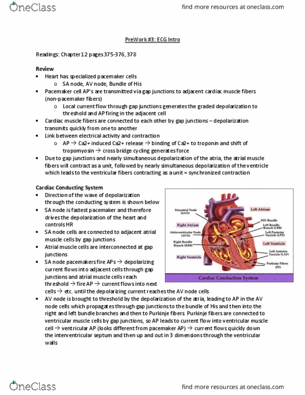
Anatomy of heart: depolarization of nodal/pacemaker cells start electrical event in heart (autorrhythmicity). If sa node damaged, av node takes over at slower rate (40bpm). Fibrous skeletons separate the chambers, so no gap junctions. Electrical signal can only go from atria to ventricle through av node. Impedance mismatch: fibers in av node are tiny compared to fibers in bundle of his. Depolarization may start in septal cells (cardiac contractile cells), but depolarization of cardiac contractile cells mostly starts at bottom of heart. Since outflow vessels on top, depolarization and contraction starting at the bottom allows blood to move to outflow vessels. Ecg: extracellular recording of electrical activity of cardiac contractile cells in heart (can"t measure nodal cells accurately because only makes up 1% of the heart: attach electrodes to arms and leg and measure electrical signal. Since humans mostly made of water and electrolytes, easy to detect electrical signal: use frontal (limb) leads and chest leads to measure ecg.