NURS 3444 Lecture Notes - Lecture 25: Anomalous Pulmonary Venous Connection, Tricuspid Atresia, Pulmonary Atresia

Document Summary
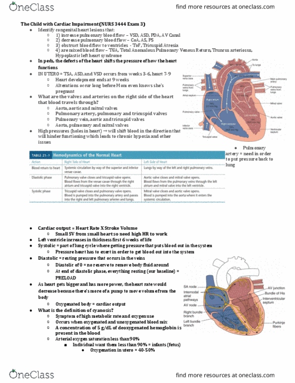
The right ventricle has a lower pressure during systole than the left ventricle because less pressure is needed to pump blood to the lungs than to the rest of the body: comparison of chd classification systems. The laboratory finding that would be seen in the cyanotic heart disease client but not in the acyanotic heart disease client would be a(an): elevated po2, elevated hemoglobin, decreased hematocrit, decreased pco2. Causes desaturated blood to shunt to the left and into systemic circulation. Usually hypoxemic and usually cyanotic: most common defects are tet and tricuspid atresia. Pulmonary or tricuspid atresia: pulmonary atresia, kids have foramen ovale so things come in atrium and try to shift over into left atrium, not getting much oxygenated blood, if you don"t have tricuspid valve. Relative desaturation of blood in systemic blood flow: cardiac output decreases because of volume load on ventricle, signs of desats, cyanosis, and chf, but variable depending on anatomy.