RIU 332 Lecture Notes - Lecture 6: Internal Obturator Muscle, Pelvic Brim, Labia Majora

20 Feb 2020
School
Department
Course
Professor
Document Summary
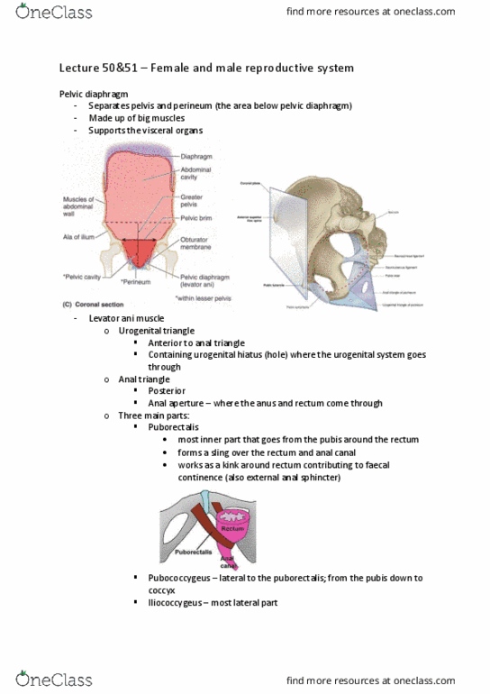
Normal anatomy and physiology of the female pelvis. Consists of four bones: two innominate (coxal) bones. Divided into two continuous compartments (true and false pelvis) by oblique plane that passes through pelvic brim muscles. Is situated inferior to caudal portion of parietal peritoneum. True pelvis is considered the pelvic cavity. Posterior wall formed by sacrum and coccyx. Posterolateral wall formed by piriformis and coccygeus. Anterolateral walls formed by hip bones and obturator. Lower margin of pelvic cavity, pelvic floor, formed by internus muscles which rim ischium and pubis levator ani and coccygeus muscles. Anterior: occupied by bladder, ureters, ovaries, fallopian tubes, uterus, vagina. Muscles extend superiorly from xiphoid process to symphysis pubis inferiorly. Base: anterior to vagina, superior surface related to uterus. Neck: rests on the upper surface of urogenital diaphragm; inferolateral surfaces relate to retropubic fat, obturator internus, levator ani muscles, pubic bone. Cross pelvic inlet anterior to bifurcation of common iliac.