Kinesiology 3222A/B Lecture Notes - Lecture 1: Axon Hillock, Thoracic Cavity, Pseudounipolar Neuron

20 Apr 2021
School
Department
Course
Professor
Document Summary
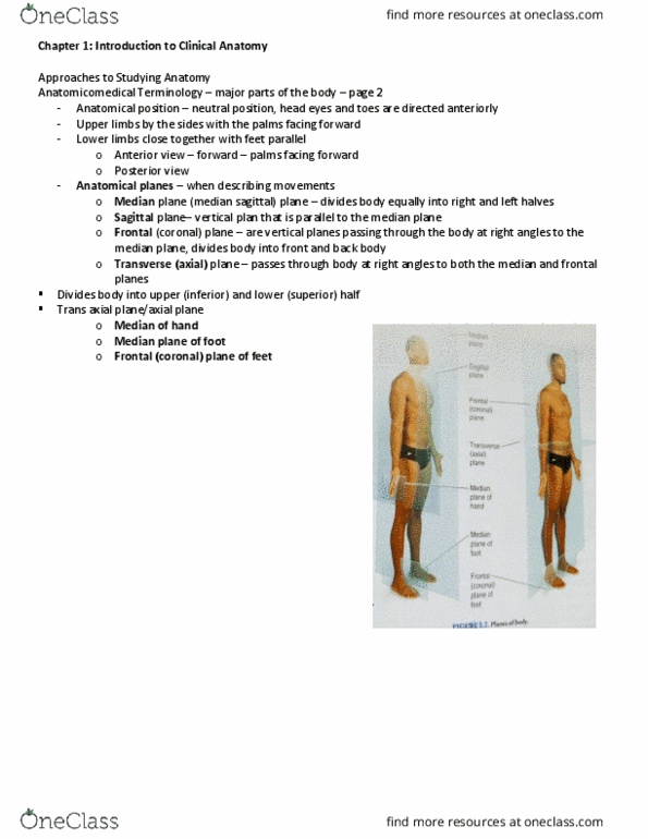
Anatomical section sagittal plane - cut body left and right halves median - goes through nose and belly button. Anatomical terms anterior- posterior radiograph of elbow arm extended in anatomical position, image shot front to back scout line: image taken in coronal plane, scout line cuts it in a separate plane. Specific terms used to denote a specific location in the body. Important for establishing a common language amongst a team. All terms are in reference to anatomical proximal: closer to midline. Week 1 notes1 distal: further away from midline. Week 1 notes2anatomical cavities cranial cavity - brain vertebral canal - spinal cord thoracic cavity - subdivided: pericardial cavity is right in the center and holds the heart, called center area area above it called mediastinum. Week 1 notes2 thoracic cavity divided from the abdominal cavity by the diaphragm pleural cavities left and right which have left and right lungs abdominal and pelvic cavities divided at pelvis.