Anatomy and Cell Biology 3319 Lecture Notes - Lecture 5: Basal Ganglia, Auditory Cortex, Cerebral Cortex

1 May 2018
School
Department
Professor

Lecture 005: Basal Ganglia
Cerebrum Review
● White matter (3 types)
○ Projection: Cerebral cortex to the rest of the brain (subcortical)
○ Commissural: Connections from hemisphere to the other (corpus callosum)
○ Association: Connections within a hemisphere
● Deep grey matter
○ Basal ganglia
■ “Ganglia” usually refers to a collections of neurons in the periphery
■ Only exception is the basal ganglia
■ Which is a large collection of neurons within the brain
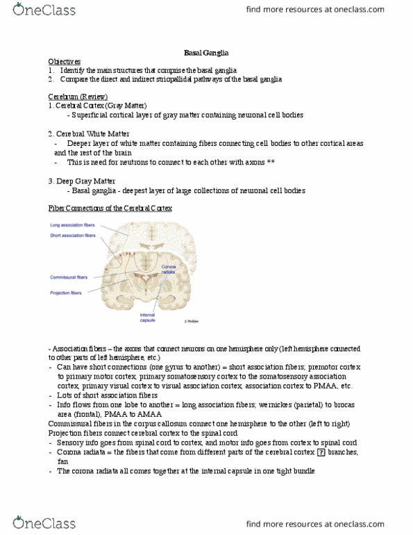
Fiber Connections of the Cerebral Cortex
● Commissural fibers
○ Largest collection is the corpus
callosum
● Projection fibers
○ Subcortical connections (how the
cerebral cortex connects to the rest of
the brain)
○ 2 types of fibers
■ Project in: Sensory information
(and other information) has to
come in
■ Project out: Motor information
has to go out
○ Blunt dissection will let you see a fan-
shaped array of fibers
■ Projection fibers come from all parts of the cerebral cortex
■ Start off broadly but they all have to connect to the internal capsule
■ Ends up with the corona radiata (fan shaped)
● Association Fibers
○ Critical for integration and processing
○ Short association fibers
■ Goes from one gyrus to another
■ Stays in one lobe and hemisphere
■ Ex. primary auditory cortex into the auditory association cortex
○ Long association fibers
■ Goes from lobe to lobe
■ Wernicke's area to Broca's area
■ PMAA to AMAA
Function of the Basal Ganglia
find more resources at oneclass.com
find more resources at oneclass.com

● Motor: coordination speed and strength of movements
● Cognition
● Learning
● Emotions
Deep Grey Matter
● Basal Ganglia
● This is a more posterior coronal section
○ Can’t see the diencephalon (no
thalamus/hypothalamus)
● Basal Ganglia also grows in a c-shaped
Basal Ganglia: glass brain view
● 3D view of the basal ganglia by pretending the
cortex is clear
● Lentiform nucleus
○ In a basal form
○ Composed of the putamen and the globus pallidus
● Caudate Nucleus
○ Forms a C-shape
■ Head, body, tail (in the temporal lobe)
● Corpus striatum
○ Lentiform + caudate nucleus
○ The neuronal bodies have fibers passing through
■ Looks striated (have lines)
● Striatum
○ Not the same as the corpus striatum
○ Caudate nucleus + putamen
● Thalamus
○ Medial to the lentiform nucleus
● Some parts of the diencephalon are clinical part of the basal ganglia
○ Subthalamic nucleus (diencephalon)
○ Substantia nigra (brain stem)
find more resources at oneclass.com
find more resources at oneclass.com
Document Summary
Projection: cerebral cortex to the rest of the brain (subcortical) Commissural: connections from hemisphere to the other (corpus callosum) Ganglia usually refers to a collections of neurons in the periphery. Which is a large collection of neurons within the brain. Subcortical connections (how the cerebral cortex connects to the rest of the brain) Project in: sensory information (and other information) has to come in. Project out: motor information has to go out. Blunt dissection will let you see a fan- shaped array of fibers. Projection fibers come from all parts of the cerebral cortex. Start off broadly but they all have to connect to the internal capsule. Ends up with the corona radiata (fan shaped) Ex. primary auditory cortex into the auditory association cortex. Motor: coordination speed and strength of movements. This is a more posterior coronal section. Basal ganglia also grows in a c-shaped. 3d view of the basal ganglia by pretending the cortex is clear.