Anatomy and Cell Biology 3319 Lecture Notes - Lecture 7: Middle Cerebellar Peduncle, Cerebellar Peduncle, Cerebral Peduncle

1 May 2018
School
Department
Professor

Lecture 007: Brainstem and Cerebellum
Brain stem is “inbetween” many structures
and the spinal cord
● Thus there will be lots of fibers pathways
running through it
○ Connect the brainstem to the
forebrain
● Cranial nerves are also connected to the
brain stem
○ Require nuclei to control them
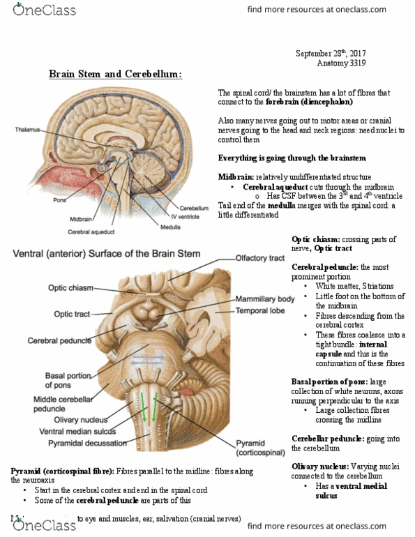
Midbrain
● Relatively undifferentiated state
● Cerebral aqueduct cuts through it
● Cerebral aqueduct connects the 3rd and
4th ventricles
Pons
● Big bulbous mass
Medulla
● Little bit of differentiation
● Merges into the spinal
cord
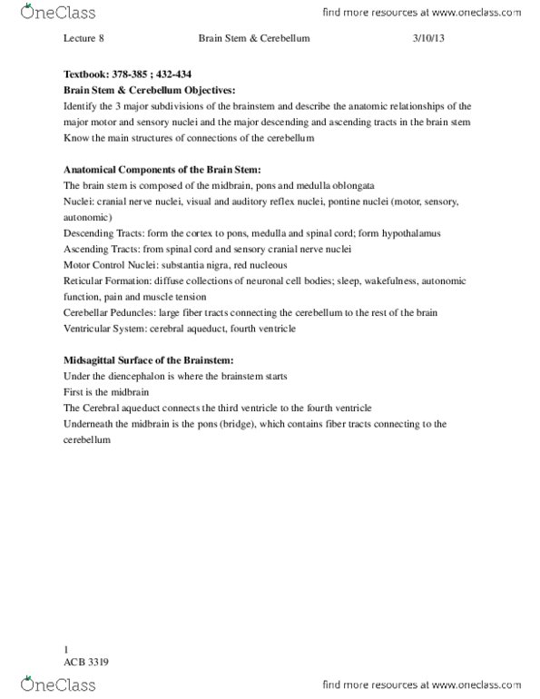
Ventral (anterior) Surface
of the Brain Stem
●Cerebral peduncle
○White matter
○ Has striation
○ Like a “foot” right
on the end of the
midbrain
○ Has relationship to the cerebral cortex
■ Motor fibers descend from the cerebral cortex
○ Continuation of the internal capsule fiber
●Basal Portion of the Pons
○ A large collection of axon fibers are running perpendicular to the neural axis
○ Continues into the Middle Cerebellar peduncle
○Middle Cerebellar peduncle
■ Large collection of fibers running crossing at the midline
●Medulla
○Oliveray nuclei
find more resources at oneclass.com
find more resources at oneclass.com

■ Connected to the cerebellum
○ Has a sulcus between the medulla and pons
○ Towards the caudal end there is a collection of fibers (pyramidal tract) crossing
the midline
■Pyramidal decussation
○ These fibers are also seen in the pyramid
■ Fibers that run parallel to the neural axis
■ These are corticospinal fibers
■ Start in the motor cortex and reaches into end in the spinal cord
●Recall: cerebral peduncle
○ Fibers that fiber that came from the cerebral cortex
○ Some of the cerebral peduncle are corticospinal fibers
● Cranial nerves run along the ventral surface of the brain stem
○ Sensory,
mortar, mix
○ Eyes, head,
muscles,
salivation,
ears
○ 12 of them
all together
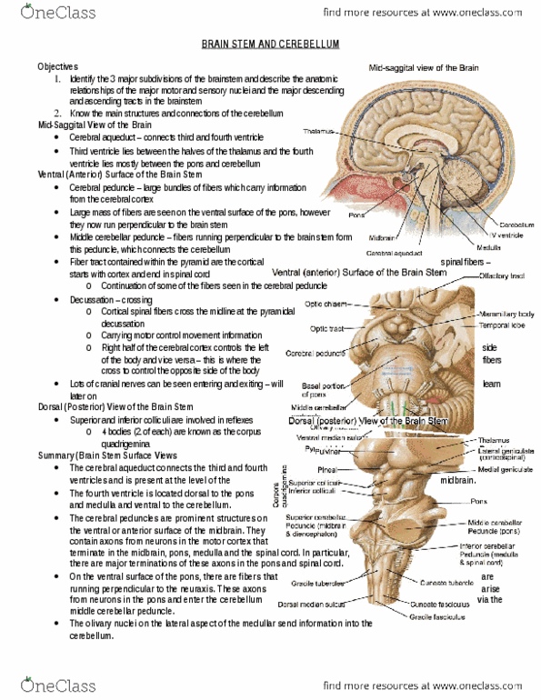
Dorsal (posterior)
View of the Brain
Stem
●Pulvinar nuclei
○ Of the
thalamus
●Lateral and
medial geniculate
nuclei
○ Auditory
and visual information
●Corpora quadrigemina (near the midbrain)
○Superior colliculi
■ Visual reflexes to track objects
○Inferior colliculi
■ Auditory reflexes to respond /orient to sounds
● Know that visual information has to travel through the thalamus then to the cerebral
cortex to “see” something
○ However, sometimes you need to do fast movements thus do not have the time
to wait for the information to travel all the way to the cortex
○ Thus the information is only passed to the midbrain and movements are
generated (reflexes - don’t need to think about it)
find more resources at oneclass.com
find more resources at oneclass.com

●Pons
○Middle Cerebellar peduncle
■ Fibers on the ventral surface cross the midline
■ Local connections (pons)
○Superior Cerebellar peduncle
■ Making connects with the midbrain (anterior regions) and the ventral
lateral regions of the thalamus (diencephalon) and cerebellum
○Inferior Cerebellar peduncle
■ Make caudal connects with the medulla and spinal cord
○ Will see more of the Cerebellar peduncle in the motor and sensory state
●Medulla
○ 2 large protrusions on the dorsal surface
○Gracile and Cuneate tubercle (bumps)
■Large sensory relay for tactile and proprioceptive sensory information
from the spinal cord that terminates within the medulla
■ Carried by the axon pathways: Gracile and cuneate fasciculus
○ Thus you will see many large fibers you if cut the medulla open
■gracile and cuneate fasciculi
● fibres bundles bring the sensory information from the spinal cord
to the gracile and cuneate tubercle
Reticular formation
● a large collection of neurons scattered throughout the brain stem
● does not form discrete nucleus
○ Raphe nucleus, medial nuclear groups, lateral nuclear groups
● control of movement, modulation of pain, autonomic reflexes and
arousal (HR, BP, breathing)
find more resources at oneclass.com
find more resources at oneclass.com
Document Summary
Brain stem is inbetween many structures and the spinal cord. Thus there will be lots of fibers pathways running through it. Cranial nerves are also connected to the brain stem. Like a foot right on the end of the midbrain. Motor fibers descend from the cerebral cortex. A large collection of axon fibers are running perpendicular to the neural axis. Large collection of fibers running crossing at the midline. Has a sulcus between the medulla and pons. Towards the caudal end there is a collection of fibers (pyramidal tract) crossing the midline. These fibers are also seen in the pyramid. Fibers that run parallel to the neural axis. Start in the motor cortex and reaches into end in the spinal cord. Fibers that fiber that came from the cerebral cortex. Some of the cerebral peduncle are corticospinal fibers. Cranial nerves run along the ventral surface of the brain stem. Auditory reflexes to respond /orient to sounds.