Anatomy and Cell Biology 3319 Lecture Notes - Lecture 8: Conus Medullaris, Cervical Vertebrae, Spinal Nerve

1 May 2018
School
Department
Professor

Lecture 008: Spinal Cord
Not very different from the developing neural tube
The spinal cord has a unique position
● Need to communicate the information of the body up
to the brain and vise versa
Injury of the spinal cord
● Loss of control of the limbs
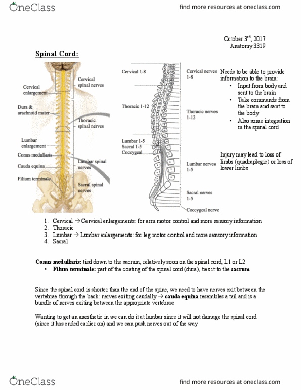
Cervical and lumbar enlargements
● Why the larger section of spinal cord?
○ More axons and cell bodies
○ For control and sensory of the limbs
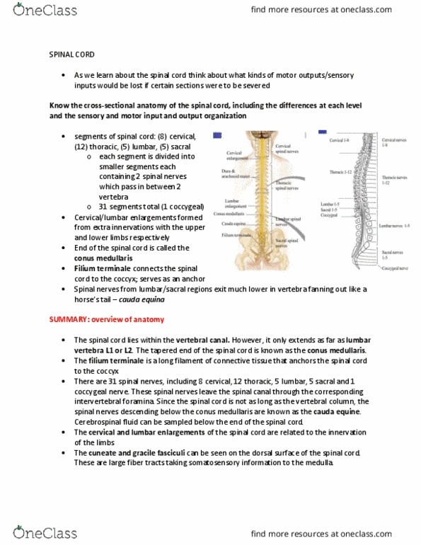
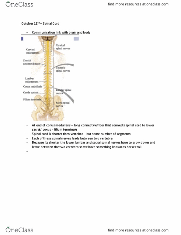
Conus Medullaris
● End of the spinal cord (a point)
● Seems to end before the end of the vertebra (ends at
L1-L2)
○ the spinal cord is shorter than the vertebra
○ This means that there is that some of the
spinal nerve has to continue to grow so that
they can exit at their appropriate vertebra
find more resources at oneclass.com
find more resources at oneclass.com

○ Thus there is a section below the conus
medullaris where there is no spinal cord but still
have a collection nerves growing down and
exiting
■ This is the cauda equina (looks like a
horse’s tail)
■ Good to inject drugs (anaesthesia) here
since the needle will simply push the
nerves aside to deposit the drug into the
lower spinal canal and not damage the
spinal cord
Filium Terminale
● Part of the thin coating of the spinal cord
● Ties the spinal cord to the sacrum
Cuneate and gracile fasciculi
● seen on the dorsal surface of the spinal cord.
● large fibre tracts taking somatosensory information to
the medulla.
Dorsal Surface of the spinal cord
● Cuneate and gracile fasciculus
○ Carries somatic-sensory information to the cuneate and gracile nuclei in the
medulla
●Dorsal root ganglia
○Part of the peripheral nervous system
■ Before the spinal cord
○ Sensory neurons carrying information from the skin and muscles
○Cell body is in the dorsal root ganglia, receptors on the skin/muscle, axons
terminates in the spinal cord
○ This is where the sensory information enters
○Dorsal root filaments goes to the dorsal aspect (grey matter) of the spinal
cord
○ Unipolar neurons
find more resources at oneclass.com
find more resources at oneclass.com

○Sensory only
●Grey matter
○ Central part
○ H-shaped (grey
commissure)
○ Ventral, dorsal,
sometimes lateral horns
●White matter
○ Outside
○ Lateral funiculus
○ Ventral funiculus
●Ventral root ganglia
○ Motor information
leaves the ventral/basal
part of the spinal cord to
the skeletal muscle
○ Carried by the ventral
root filament
○Motor only
● In the dorsal and ventral roots
the modality are separated
(only sensory or only motor)
● These modalities join in the
mix spinal nerve
○ Dorsal and ventral root join
○ Has both sensory and motor information
find more resources at oneclass.com
find more resources at oneclass.com
Document Summary
Not very different from the developing neural tube. Need to communicate the information of the body up to the brain and vise versa. For control and sensory of the limbs. End of the spinal cord (a point) Seems to end before the end of the vertebra (ends at. The spinal cord is shorter than the vertebra. This means that there is that some of the spinal nerve has to continue to grow so that they can exit at their appropriate vertebra. Thus there is a section below the conus medullaris where there is no spinal cord but still have a collection nerves growing down and exiting. This is the cauda equina (looks like a horse"s tail) Good to inject drugs (anaesthesia) here since the needle will simply push the nerves aside to deposit the drug into the lower spinal canal and not damage the spinal cord. Part of the thin coating of the spinal cord.