Anatomy and Cell Biology 3309 Lecture Notes - Lecture 9: Dict, Loose Connective Tissue, Esophagus

29 Apr 2021
School
Department
Professor
Document Summary
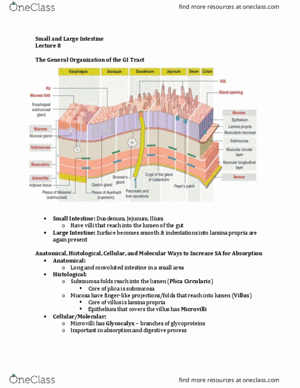
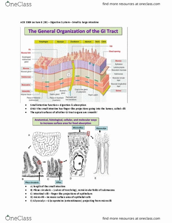
Esophagus and stomach both have a smooth mucosal surface. This changes when we reach the small intestine - they have villi. The large intestine lacks these villi; it looks very similar to the stomach. Anatomically: small intestine is a long tube with longitudinal and circumferential folds in the walls - plica circularis. Circularis because they run circumferentially around the small intestine. The mucosa of each plica circularis has intestinal villi - stick up into the lumen. Each villi is covered in an epithelium - the epithelial cells are called enterocytes - and each enterocytes has microvilli. Enterocytes are absorptive cells - they function to absorb nutrients. Microvilli are cellular structures, villi are multicellular structures. The microvilli have surface modifications called the glycocalyx - glycosylated proteins with a particular functions. Plica circularis all over intestinal walls - they are folds. They contain in its centre, a core of submucosa (the blue in the image)