NURS 1750 Lecture Notes - Lecture 9: Cranial Cavity, Bipolar Disorder, Vestibular System

28 Jun 2018
School
Department
Course
Professor

Week Seven (October 30 - November 3, 2017)
Anatomy and Physiology
Principal Parts of the Brain
- The four principal parts of the brain are the brainstem, cerebellum, diencephalon, and
cerebrum (largest part of brain).
Protection - What
protects our brain?
-Cranial bones.
-Cranial meninges.
- Pia (inner layer).
- Arachnoid (middle layer).
- Dura mater (outer 2 layers periosteal layer (external) & meningeal layer
(internal).
-Cerebrospinal fluid.
- Protects from chemical and physical harm.
- Carries oxygen, glucose and other substances from blood to nervous tissue.
find more resources at oneclass.com
find more resources at oneclass.com

Week Seven (October 30 - November 3, 2017)
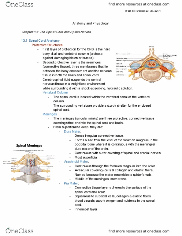
Meninges - protective coverings of the brain.
→ Cranial meninges continuous with the spinal meninges,
What are the meningeal layers called?
- Dura mater, Arachnoid mater, Pia mater
- Cranial dura mater has two layers- periosteal and meningeal
- Spinal dura mater one layer
Meninges in the Brain -- Sinuses
- Venous sinuses-endothelial-lined
venous channels-drain venous blood
from brain deliver into the internal
jugular veins.
- Cranial dura mater layers are fused except at sinuses-
drains venous blood into internal jugular veins.
Meninges in Brain
- No epidural space around brain.
- Blood vessels that enter brain tissue pass along the
surface of brain and as they penetrate inward, they are
sheathed by a loose-fitting pia mater.
Membranes
- Three extensions of the dura mater separate parts of the brain (hemispheres):
- Falx Cerebri: separates the two hemispheres (sides) of the cerebrum.
- Falx Cerebelli: separates the two hemispheres of the cerebellum.
- Tentorium Cerebelli: separates the cerebrum from the cerebellum.
find more resources at oneclass.com
find more resources at oneclass.com

Week Seven (October 30 - November 3, 2017)
Blood Flow to the Brain
- Blood flow to the brain is mainly via the vertebral and (internal) carotid arteries; the dural
venous sinuses drain the internal jugular veins to return blood from the head to the heart
(in other words, and flows back to the heart via the jugular veins)
Importance of Blood Flow to the Brain
- 2% of total body weight in an adult.
- Consumes 20% of body’s oxygen supply.
- Neurons use ATP (from glucose/oxygen).
- Increased brain activity causes increase in blood flow. Brief slow of brain blood
flow may cause disorientation or Loss Of Consciousness (LOC).
- 1-2 minutes of blood flow interruption impairs neural function.
- 4 minutes of blood flow interruption causes permanent injury.
- Glucose:
- Flow must be continuous-not stored in brain cells (brain does not
have/make glucose)
- Deficiency of glucose may produce mental confusion, dizziness,
convulsions, and unconsciousness.
- People with diabetes must be vigilant about their blood sugar levels
because these levels can drop quickly, leading to diabetic shock, which is
characterized by seizure, coma, and possibly death.
find more resources at oneclass.com
find more resources at oneclass.com
Document Summary
The four principal parts of the brain are the brainstem, cerebellum, diencephalon, and cerebrum (largest part of brain). Dura mater (outer 2 layers periosteal layer (external) & meningeal layer (internal). Carries oxygen, glucose and other substances from blood to nervous tissue. Cranial meninges continuous with the spinal meninges, Cranial dura mater has two layers- periosteal and meningeal. Venous sinuses-endothelial-lined venous channels-drain venous blood from brain deliver into the internal jugular veins. Cranial dura mater layers are fused except at sinuses- drains venous blood into internal jugular veins. Blood vessels that enter brain tissue pass along the surface of brain and as they penetrate inward, they are sheathed by a loose-fitting pia mater. Three extensions of the dura mater separate parts of the brain (hemispheres): Falx cerebri: separates the two hemispheres (sides) of the cerebrum. Falx cerebelli: separates the two hemispheres of the cerebellum. Tentorium cerebelli: separates the cerebrum from the cerebellum. 2% of total body weight in an adult.