PCL102H1 Lecture Notes - Lecture 16: Drug Design, Hiv-1 Protease, X-Ray Crystallography

27 Apr 2016
School
Department
Course
Professor

Document Summary
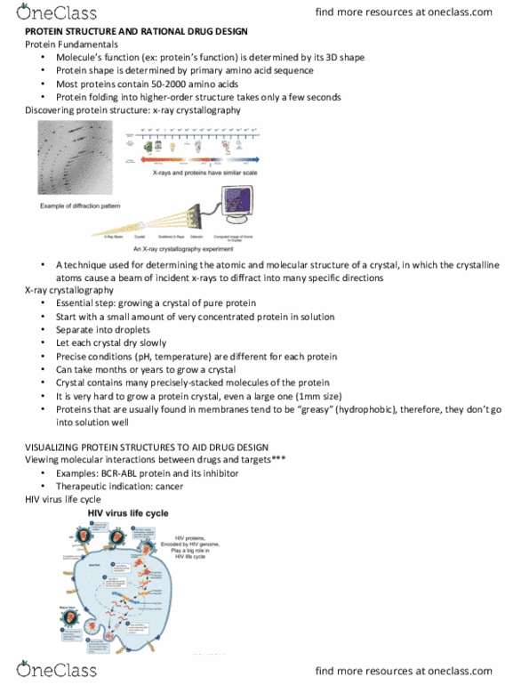
X-ray crystallography shine x-ray at crystal to see shadow of protein, use detector to see patterns to determine what crystal looks like x rays are best used for small molecules like proteins steps. Start with small amount of very concentrated protein in solution, separate into droplets, let each crystal dry slowly. Crystal contains precisely packed molecules of the protein. Very hard to grow a protein crystal, even large one (1 mm) Proteins found in membranes are hydrophobic and don"t go into solution well. Hiv protease symmetrical structure (two protease molecules form the active enzyme) first drug design. Take structure of natural substrate, cut in half, rotate 180 deg, glue two halves back together lead compound inhibited enzyme, but not water soluble so couldn"t be drug drug-resistant versions of hiv protease. Screen drugs virtually against all mutant forms of enzyme. Mutant version: up to 2000x less binding between drug and mutant enzyme.