PSY290H5 Lecture 3: PSY290 Lecture 3


PSY290
Lecture 3
Neurophysiology: The Generation, Transmission, and Integration of Neural Signal
Last lecture: macro anatomy of the brain (seen with the naked eye)
This lecture: microanatomy that exists within the brain
A tour of the parts of the micro anatomy of the brain
Neuroanatomy
Neurons
Neurons are more than the classic shape that we are familiar with
There are hundreds of different types of neurons and those serve a variety of functions
oSame as the brain layer architecture (different emphasis on layers mean different functions)
Different emphasis on neuron features mean different functions
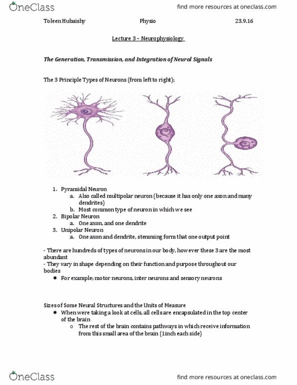
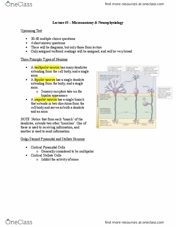
Multipolar neuron: reaches out for a lot of input and sends out 1 output from it
oFunction is most commonly associated with nerves in our body that receive and send
information over a long range
Cyadic nerve: base of spine all our way to our leg and big toe
Bipolar neuron: the distinction between the parts is not quite evident
oMost commonly associated with sensory type neurons
oTouch, sight, auditory
oReceive, process, and send alone over a much shorter distance
oFewer connections with things
oNot as multiconnected as a multipolar neuron is
Unipolar neuron: a cell body with a single extension
oOne zone of information
oBranches extend away from the cell body to form an input zone and an output zone
oUnipolar neurons are most associated with some kind of interneuron function
Any kind of neuron that connects other neurons with each other that links information
across the brain
Golgi-Stained
Left hand side: cortical pyramidal neuron
oOne big branch and connected to the cell body with branches out of it
o80% of neurons of the brain
oresponsible for excitatory information
meaning: connecting, conveying, sending information from one neuron to another
Right hand side: Cortical Stellate neuron
oDoes not have any huge branch extending
oOnly a bunch of little branches connected to it
oDoes not fit with the 3 definitions mentioned above
oStellate cells are well known to perform inhibitory functions in the brain
prevent further activity of neurons
the brain needs both excitation and inhibition to work properly
like driving a car: we need accelerator and a brake
Question: what type of neuron is a cortical pyramidal neuron? Multipolar
*know how to know type of neuron from a picture
*know the parts of the neurons from a picture

Parts of the Neuron
4functional zones associated with neurons
Cell Body:
contains all the functional parts/machinery required for the cell to work
omitochondria
oorganelles
onecessary to produce proteins for translation and transcription
parts of the neuron that collects and integrates incoming information
otakes electrical signals and it processes them in a simplistic kind of way
oinvolved in processing input information
found inside of the input zone: dendrites
oreceive information from other neurons
othe greater the surface area (more branches), the more connections dendrites can make with
other neurons and more information they can integrate
ointegration of information can be bad or good: the more integration, the more acviity
generated (more sensitivity) but the vaster the information (more space covered), the less the
accuracy
oknow the pros and cons of integration
obranches that exist on dendrites
dots that extend from dendrites
when we think about synaptic plasticity and learning memory on the level of the
neuron
when kids develop, the difference is not the number of cell bodies, not number of
axons, but what does change dramatically with learning are the dendrites
dendrites are extremely important for synaptic plasticity: the ability of neurons to
change with experience
dendrites do 2 things
receive information from other neurons
change with experience (learning on the neuronal level)
Axons
oDo not change often
oFixed in location
oVery long branch that extends away from the cell body
oAxons are the primary transmission line of the nervous system
oDo not represent any integration of input, but rather the transmission of information to other
places
oConducting zone in terms of functional zones
oThe action potential: how axons transmit information
oIndividual axons are generally very tiny
Cyadic axon is the longest one
Can be over a meter in length
oFunctionally, the axon of a neuron has interesting functional properties
Neuronal membranes have special connection areas: ion channels

Proteins that span some aspect of the surface of a cell (the neuron)
These channels allow ions to pass through it
The way that neurons work is that they have a special property about ion
enhance that neurons can actually hold an electrical charge by a balance
between the inside and outside of a neuron: similar to a battery
The neuron is not the only tissue that creates this electrical balance
oThe muscles surrounding the heart also have an electrical charge to
induce change
Selective Permeability
Ion channels are associated with a passive property
Axons are lined with special kinds of ion channels: voltage-dependant channels
oTo open and close, they require an electrical change in the conductivity of the cell at one
moment
Ligand Gated Channels: channels that require a ligand
oA ligand is any chemical that causes a biological interaction (drugs, hormones,
neurotransmitters)
Sodium-Potassium Pump: a pump that requires energy
Calcium Channel
Cytoskeleton
Axons require structural support
That support helps to take all the product from cell body and helps send it to where it needs to be
4 types of structural support
omyelin
omicrotubule
oneurofilament
omicrofilament
proteins are carried down these tracks
these 4 structures are along the axon
all these filaments are actively moving elements (proteins or others) down an axon to where it needs
to be
these filaments are so important to neural function and if any is affected, a signal is affected
filaments are tightly wrapped together with a string with a tao
oin a healthy brain, tao connects and maintains structural order to the 4 filaments
oin Alzheimer’s disease, for some reason, the amount of string, tao, starts to increase in the
brain
those phosphate molecules sense too much tao and orders them to be cut off
this signal starts to cut too many tao proteins and as a result, the tao becomes less able
to hold microfilaments together
those filaments start to become unwound or loosen from one another
the result is a disorganization of neurons inside of the brain
top box: healthy area little better organization or axons and neurons
below box: evidence of disorganization in the brain
beginning of losing more and more neurons
Alzheimer’s Disease
we only confirm diagnosis after death
axons tend to lose their shape and structure: Tangles