BCH 4101 Lecture Notes - Lecture 3: Polyacrylamide Gel Electrophoresis, Genome Project, Human Genome Project

28 Apr 2018
School
Department
Course
Professor

September 20, 2017
The Human Genome Sequencing Project
Sequencing: the Beginning
1953: 3D structure of DNA is elucidated
-Using Rosalind Franklin and Maurice Wilkins’ X-ray crystallography data, James Watson and Francis Crick were
able to elucidate the structure of DNA
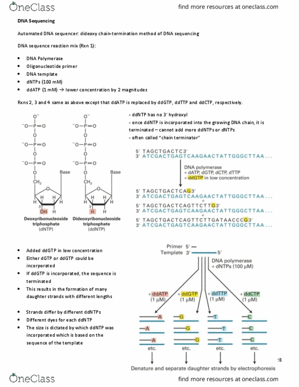
Mid 1970’s: Frederick Sanger develops the “Sanger” method of DNA sequencing (aka chain-termination method,
dideoxy sequencing)
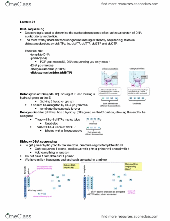
-Adding a ddNTP instead of a dNTP during DNA synthesis stops chain elongation at that point because the 3’-OH is
required for the creation of a phosphodiester bond with the 5’ phosphate of the next nucleotide
•ddNTPs = chain terminators
-Use radioactively labelled ddNTPs and set up four different reactions with a ssDNA template, DNAP, dNTPs, and a
trace amount of ddNTPS
•Set up a reaction with ddATP, another reaction with ddTTP, another with ddCTP, and another with ddGTP
•Need four reactions because radioactivity doesn’t have different colours
-You can then separate the fragments using polyacrylamide gel electrophoresis and read the sequence manually
•Each well represents one of the four reactions
•Order the fragments based on length
•Fragments on the bottom of the gel (shorter) represent the beginning of the sequence, fragments at the top of
the gel (longer fragments) represent the end of the sequence
•From this sequence, you can deduce the sequence of your original sequence
-The sequence you get is complimentary to your original sample
•NB: need large gels with high percent of acrylamide in order to get proper separation between fragments
-1977: phage phiX174 (5.3 kb) genome was sequenced
-1982: phage lambda (48 kb) genome was sequenced
-Disadvantages: very labour intensive and time consuming
•Manual process
•Can only read ~50-100 nucleotide sequences
1986: Leroy Hood (Caltech) invented fluorescent ddNTPs and allowed the sequencing process to become automated
-Used four different fluorescent dNTPs
-Now only need one reaction - all reactions performed in the same tube
1

September 20, 2017
-Use capillary gel electrophoresis to separate the fragments and measure fluorescence as the fragments leave the
tube
•Allows for better separation
-Advantages:
•96 or 384-well plates can be sequenced simultaneously (3 hours) by using the capillary-sequencing method
•Can now read ~500-800 bases
•Cost reduction: $3/read vs. $30-$100/read with the manual method
•Automated: use of robots
-Automated sequencing and capillary electrophoresis pushed the technology far enough that genome sequencing
projects were completed ahead of schedule and under budget
•Revolutionary turning point
1986: United States Department of Energy is seeking data on protecting the genome from the mutagenic effects of
radiation
1988-89: US Government funds the National Centre for Human Genome Research (led by James Watson)
-Goal was to “coordinate research and technical activities related to the human genome”
1988: scientists propose to sequence the human genome
-Challenges:
•Automated sequencing still isn’t reliable enough
•Availability of clones to be sequence
-Didn’t have enough clones to cover the entire genome
-Didn’t have enough stuff to sequence
•Assembly of the complex sequence
-How do you know which pieces are supposed to go together?
1990: International Human Genome Sequencing Consortium (IHGSC) is formed
-Initial goal: sequence the euchromatin portion of the genome (2.85 x 10⁹ bp = 92% of the genome)
•Do it in 15 years on a budget of 3 billion dollars
-Collaborative effort among more than 26 countries
Human Genome Project
Goals of the project:
-Construction of a high-resolution genetic map of the human genome
2

September 20, 2017
-Production of a variety of physical maps of all human chromosomes and of the DNA of selected model organisms,
with emphasis on maps that make the DNA accessible to investigators for further analysis
-Determination of the complete sequence of human DNA and of the DNA of selected model organisms
-Development of capabilities for collecting, storing, distributing, and analyzing the data produced
-Creation of appropriate technologies necessary to achieve these objectives
Initial timeline:
-Phase 1 (1990-1997):
•Create detailed genetic and physical maps
•Sequencing of simple genomes to test the technology
-Phase 2 (1997-2001):
•Sequencing of the human genome (gaps and errors)
-Phase 3 (2001-2003):
•Finishing of the sequence
•<1 error per 104 bp
•Permanent foundation for all human biology and biomedical research
J. Craig. Venter:
-Recruited by the NIH in 1984
-Early 1990s: developed the EST method of finding genes in the brain
-Tried to patent the genes he identified in the brain, but NIH abandoned the applications after huge public outcry
-Venter quit the NIH and started his own non-profit, the Institute for Genomic Research (TIGR) in 1992
-Developed a quicker/faster method of sequencing (whole genome shotgun sequencing)
-Applied for an NIH grant for sequencing of H. Influenza genome
-Started without waiting for NIH grant and was able to almost complete the sequence when the NIH rejected the
proposal on the basis that it wouldn’t work
•Completed the sequence in 1995 (1.83 Mbp)
NIH team: construction of genetic and physical maps of the human and mouse genome (anchoring points for
assembly of genomic sequence)
-Was able to complete the sequencing of smaller genomes
•1996: Yeast (12 Mb)
•1997: E. Coli (4.7 Mb)
•1998: C. Elegans (97 Mb)
3
Document Summary
Using rosalind franklin and maurice wilkins" x-ray crystallography data, james watson and francis crick were able to elucidate the structure of dna. Mid 1970"s: frederick sanger develops the sanger method of dna sequencing (aka chain-termination method, dideoxy sequencing) The sequence you get is complimentary to your original sample: nb: need large gels with high percent of acrylamide in order to get proper separation between fragments. 1977: phage phix174 (5. 3 kb) genome was sequenced. 1982: phage lambda (48 kb) genome was sequenced. Disadvantages: very labour intensive and time consuming: manual process, can only read ~50-100 nucleotide sequences. 1986: leroy hood (caltech) invented uorescent ddntps and allowed the sequencing process to become automated. Now only need one reaction - all reactions performed in the same tube. Use capillary gel electrophoresis to separate the fragments and measure uorescence as the fragments leave the tube: allows for better separation.