BPK 306 Lecture Notes - Lecture 29: Optic Radiation, Retinotopy, Peripheral Vision

5 Dec 2017
School
Department
Course
Professor
Document Summary
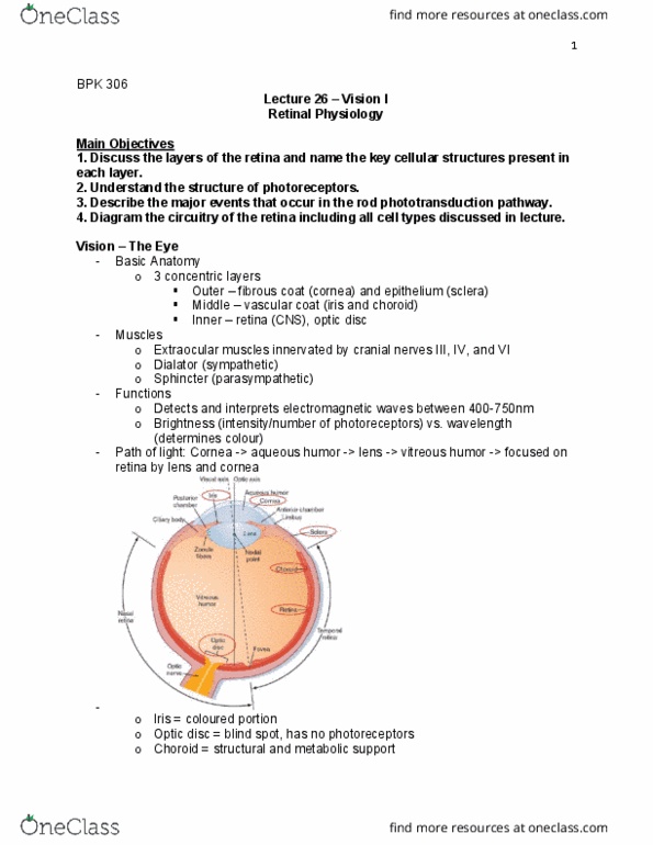
In the fovea, there is a more even ratio between prs and gcs (~1:1: small receptive field and good resolution, but low sensitivity. In the remainder of the retinal, gcs are connected to a varying number of prs (~15-45) Foveal and extrafoveal vision differ in resolution and colour perception: foveal -> colour, high acuity vision (cones, extrafoveal -> dim light, low acuity vision (rods) 2: optic chiasm -> the point where the nasal retina tracts crossover, binocular segment of target imaged on both retinas. In the primary visual cortex -> representation of space is flipped left->right and up->down. 6 layers -> each receives input from the ipsilateral or contralateral eye. Rfs of lgn cells is similar to that of corresponding gc cell. Magnocellular layers (1 and 2) have center-surround receptive fields and receive input from rods in peripheral vision (movement and brightness) Parvocellular layers (3-6) have center-surround receptive fields and receive input from cones in foveal vision (colour and form)