NSE 22A/B Lecture Notes - Lecture 8: Lactiferous Duct, Sebaceous Gland, Smooth Muscle Tissue

Document Summary
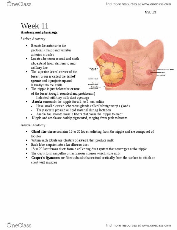
Physical examination and health assessment. (2 canadian ed. ) (canadian editors: browne, a. , Identify and describe the normal anatomy of the breast including: tail of spense, nipple, areola, cooper ligaments and adipose tissue. Superior lateral corner of breast tissue: project up and laterally into the axilla. Nipple: below the centre of the breast, rough, round, and usually protuberant. Surface looks wrinkled an indented w/ tiny milk duct openings. Inside are small elevated sebaceous glands called montgomery"s glands: secrete a protective lipid material during lactation. Smooth muscle fibers that cause nipple erection: nipple and areola are more darkly pigmented than the rest of the breast surface. Fibrous bands extending vertically from the surface to attach on chest wall muscles. Support breast tissue: become contracted in breast cancer; pits or dimples in overlying skin. Layer of subcutaneous and retromammary fat actually provide most of the bulk of the breast.