PSYCH 1XX3 Lecture Notes - Lecture 4: Wilder Penfield, Phineas Gage, Montreal Procedure

22 Feb 2017
School
Department
Course
Professor

find more resources at oneclass.com
find more resources at oneclass.com

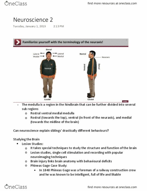
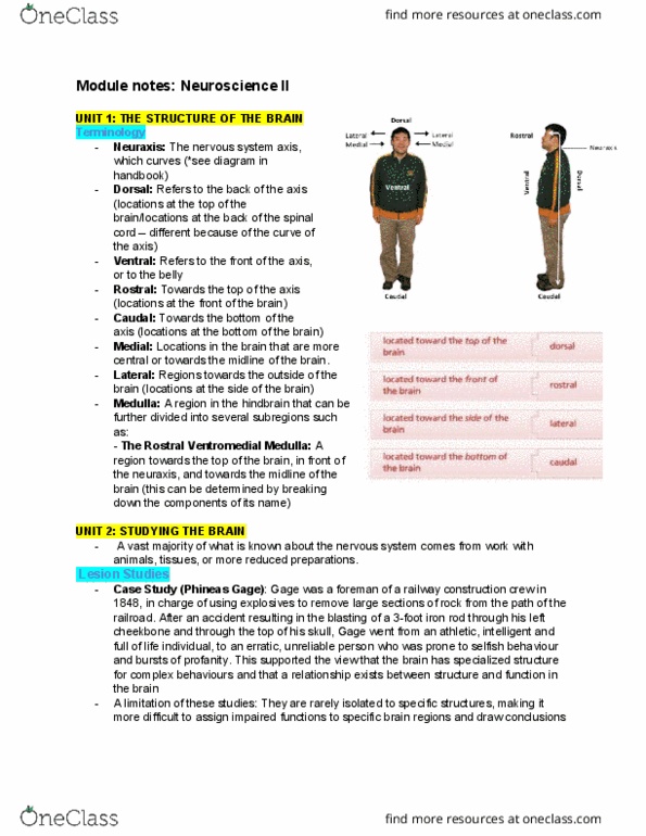
UNIT 1- THE STRUCTURE
OF THE BRAIN
Neuraxis- curves at the head
Corsal- back of axis (at the spinal cord),
and up (at the level of the head)
Ventral- front of the axis
Rostral- top of the axis
Claudial- bottom of the axis
Medial- central (towards the center of the
brain)
Lateral- towards the outside of the brain
- ie: medulla- subsection: rostral
ventromedial medulla
- Diff contributions to behaviour are
made by key brain areas
UNIT 2- STUDYING THE BRAIN
Important ways in which scientists in the past and present have made sig. advancements in the
field of neuroscience.
- Lesion studies, single cell stimulation and recording and popular neuroimaging tech.
Lesion Studies:
- How can accidental brain injury link anatomy with
associated cog and beh deficits?
- ie: Phineas Gage- got into an explosive accident on the
railroad. This changed how he acted, his personality as
well as his reliability.
- Supports the view that the brain has specialized
structures for complex brain patterns.
-Advantage: direct measure of brain’s structure and function
-Disadvantage: human brain lesions are rarely isolated to spec structures. This makes is
difficult to target particular regions and draw conclusions
- Solution: specific brain lesions can be studied in animal models
- Destroy remove or deactivate specific brain region and observe the result on beh
- But still, often a variety of beh are affected y a single lesion.
Stimulation and Single Cell Recording:
- Electrically stimulate an area of the brain and observe the results on beh (microelectrode
targeting a single neuron)
- Wilder Penfield used this and revolutionized techniques in brain surgery.
find more resources at oneclass.com
find more resources at oneclass.com

- Penfield used this procedure (the “Montreal Procedure”) to cure epileptic
seizures
- The tricky part was that he had to keep the important areas of the brain intact
and undamaged
- He used a wire to stimulate the individual cells
- In the procedure the patients can be awake, because there is no pain receptors
in the brain. This allows the patient to work alongside penfield to figure out what
different parts of the brain do.
- Electrodes can be used to record ongoing electrical activity in the brain through single
celled recording techniques
- The neural activity is recorded through a small electrode inserted into the
nervous tissue of an live animal model with its tip held just outside the cell body
of an indiv neuron
- When the neuron is firing it reveals a specific neuron’s functional role
- ie: cats were presented with visual stim and certain sections of the brain lit up
when presented with different categories of pictures
Structural and Functional Neuroimaging:
- Looks at the large-scale structure and function of brain regions. (Instead
of just the function of specific neurons)
-CT (Computed tomography)
- The first struct neuroimaging technique developed
- A series of X-ray slices of the brain are taken and pieced together
to produce a relatively quick and inexpensive picture of the brain.
- Helpful to diagnose brain injury
- Limitation:relatively low resolution (by today’s standards); difficult
to examine fine brain anatomy with a CT scan
- Inexpensive picture of the brain
-MRI (magnetic resonance imaging):
- Powerful magnetic fields are produced in an MRI machine, which align the
hydrogen atoms.
- When the hydrogen atoms are aligned, the MRI can easily localize tissue very
precisely throughout the brain.
- Precise detailed picture of the brain
-PET (Positron emission tomography)
- To learn how the brain functions in relation to cognitive tasks (ie: language,
memory.)
- A radioactive tracer of glucose or oxygen is injected into the bloodstream.
- The radioactive markers make their way to the brain and are used in metabolic
processes , which are detected by the PET scan.
- The more active the area of the brain is the more metabolic resources it will use.
- The areas that have the most radioactive tracers are the most active
areas.
- Disadvantage: invasive procedure of injecting the radioactive tracer.
find more resources at oneclass.com
find more resources at oneclass.com
Document Summary
Corsal- back of axis (at the spinal cord), and up (at the level of the head) Medial- central (towards the center of the brain) Lateral- towards the outside of the brain ie: medulla- subsection: rostral ventromedial medulla. Diff contributions to behaviour are made by key brain areas. Important ways in which scientists in the past and present have made sig. advancements in the field of neuroscience. Lesion studies, single cell stimulation and recording and popular neuroimaging tech. How can accidental brain injury link anatomy with associated cog and beh deficits? ie: phineas gage- got into an explosive accident on the railroad. This changed how he acted, his personality as well as his reliability. Supports the view that the brain has specialized structures for complex brain patterns. Advantage: direct measure of brain"s structure and function. Disadvantage: human brain lesions are rarely isolated to spec structures. This makes is difficult to target particular regions and draw conclusions.