KINESIOL 1Y03 Lecture Notes - Lecture 15: Musculocutaneous Nerve, Medulla Oblongata, Foramen Magnum

27 Dec 2014
School
Department
Course
Professor
Document Summary
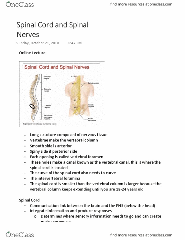
Each individual vertebra has an opening called vertebral foramen (hole). Successive holes create spinal canal: canal: several curves. Anchor spinal cord laterally, prevent from side-to-side movement: filum terminale maintaining spinal cord, anchor in place, subarachnoid space (between arachnoid mater and pia mater), contains csf, delivers nutrients, eliminates waste. Organization in the spinal cord: sensory neurons pass into posterior horn from periphery. Spinal nerves: numbered for region of vertebral column where they exit, first pair exist between skull and first cervical vertebrae, four pairs exit via the sacral foramina (bones fused together through aging, others exit through intervertebral foramina. L4-s4 = sacral plexus: s5 co= coccygeal plexus. Structure of peripheral nerves: axons bundled in parallel (motor and sensory, grouped based upon function, endoneurium axon surrounded by connective tissue layer (3, this is the first) Innervates superficial neck structures, skin of neck, posterior portion of head: contains phrenic nerve (c3-c5) which innervates diaphragm.