PSYC 212 Lecture Notes - Lecture 1: Craniotomy, David H. Hubel, Retinal Ganglion Cell

21 Apr 2016
School
Department
Course
Professor
Document Summary
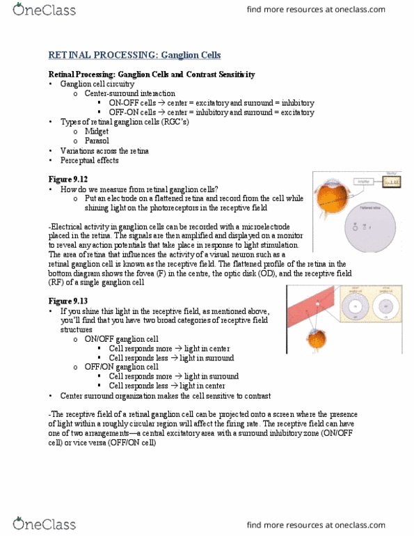
Short review of retina: division of labor, spatial. Fovea high resolution vision, poor low-light sensitivity (no rods) Periphery low-resolution vision, good low-light sensitivity (rods in periphery: photoreceptors. Rods only sensitive in low-light, bleached when exposed to too much light no color perception, rhodopsin. Cones only sensitive from medium to high-light, good for color perception, photopigment sensitive to s-, m-, l-wavelength light: ganglion cells (main output cells messages to brain) Midget cells small rf"s, color sensitivity, but poor temporal response. Parasol cells large rf, poor color sensitivity, great temporal response: processing and output, center-surround rf"s, contrast detection. The visual system: figure 10. 3: how information from the visual field is combined, retina divides into, temporal portion of retina goes through the optic nerve. At the optic chiasm it stays on the same side (ipsilateral projection: nasal portion of the retina crosses over to the other side (contralateral projection, this happens for both eyes, cross-over happens at the chiasm.