PHGY 209 Lecture Notes - Lecture 8: Retinal Ganglion Cell, Retinal Pigment Epithelium, Fovea Centralis

4 Jan 2017
School
Department
Course
Professor
Document Summary
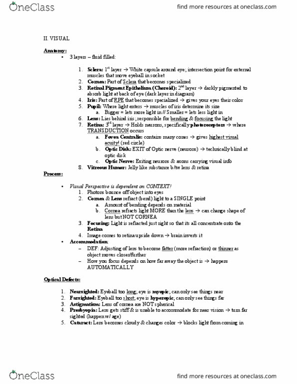
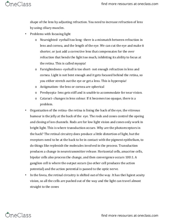
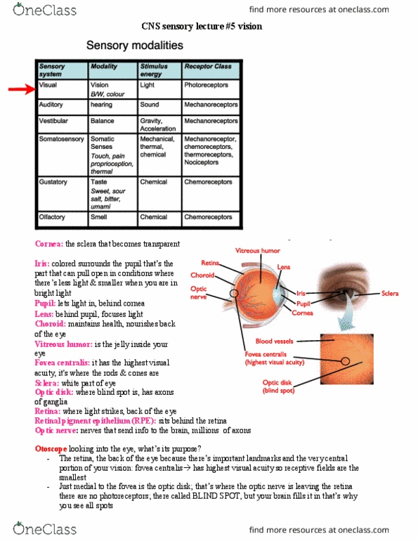
Visual sensory system: retinal pigment epithelium is other name for choroid. Iris is what forms the pupil: blood vessels are on the surface of the retina, fovea centralis is in the middle of the retina, optic disk --axons leaving through optic nerve, no photoreceptors (blind spot) Color blindness: genetic disease with problem in opsin molecule. Cortical representation of visual world: line segments, no longer donuts. Hearing loss: hearing decreases as get older (presbycusis), but exposure to loud noises accentuates the decline. Anatomy of the ear: middle ear is air-filled channel, mechanical coupling from tympanic membrane by little bones, must have same air pressure in front and behind the ear drum, popping airplanes when air rebalances (yawn opens eustachian tube) Its motion is frequency dependent: more flexible membrane towards the end, low frequency towards the end (low pitch) and high frequencies close to the beginning (high pitch)